Brain Tumor Res Treat.
2018 Oct;6(2):73-77. 10.14791/btrt.2018.6.e9.
Pineal and Suprasellar Germinoma Cooccurence with Vertebra Plana: A Case Report
- Affiliations
-
- 1Department of Radiology, Faculty of Medicine, Mashhad University of Medical Sciences, Mashhad, Iran. aminzadehb@mums.ac.ir
- 2Department of Neurosurgery, Mashhad University of Medical Sciences, Mashhad, Iran.
- 3Psychiatry and Behavioral Sciences Research Center, Mashhad University of Medical Sciences, Mashhad, Iran.
- KMID: 2423977
- DOI: http://doi.org/10.14791/btrt.2018.6.e9
Abstract
- Germinoma is the most common type of intracranial germ cell tumors (GCTs). Pineal gland and suprasellar region are the most frequent sites of central nervous system (CNS) involvement. Intracranial masses caused by Langerhans cell histiocytosis (LCH) mimics features of CNS GCTs. LCH frequently involve spine and is the most common cause of vertebra plana in children. A 15-year-old boy presented with progressing symptoms of polydipsia, polyuria, general headache, nausea and severe back pain. Brain MRI showed brain tumor with simultaneous involvement of suprasellar region and pineal gland. An excisional biopsy of suprasellar mass was done. The pathologic assessment confirmed the diagnosis of germinoma. Patient's treatment continued accordingly. A spine MRI, done due to persistent backache, showed a vertebra plana. We reevaluated the primary diagnosis suspecting LCH. Germinoma of CNS was confirmed and a biopsy of vertebral lesion resulted in hemangioma. Thus we report a case of CNS germinoma with co-occurrence of vertebra plana. We emphasized the importance of histopathologic diagnosis of pineal/suprasellar masses and primary investigation of other CNS regions including spine for possible metastasis or comorbidities.
Keyword
MeSH Terms
Figure
-
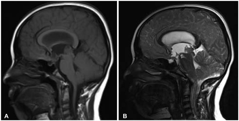
Fig. 1 Sagittal T1-weighted (A) and T2-weighted (B) images show two complex solid-cystic masses, a 17 mm mass located in pineal region and a 15 mm mass in suprasellar region.
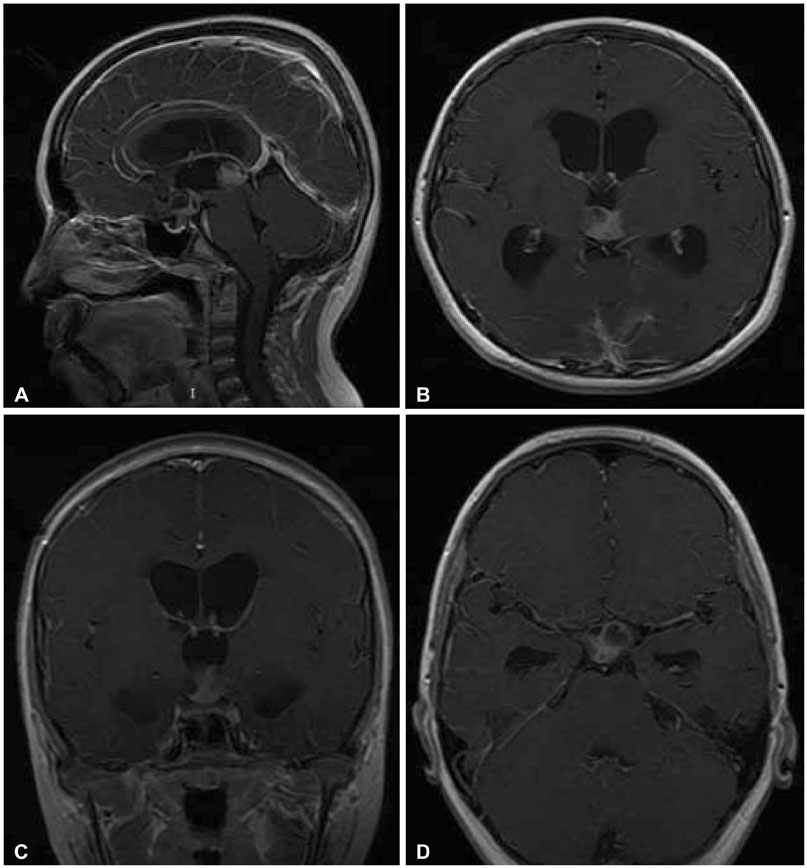
Fig. 2 Sagital (A), axial (B and D), and coronal (C) post contrast T1-weighted images show enhancement in solid parts in both masses.
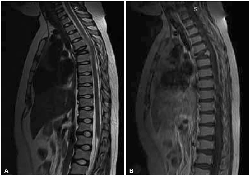
Fig. 3 Sagital T2-weighted (A) and T1-weighted (B) images shows complete collapse of T5 vertebral body (vertebra plana) with preservation of disk space. Moderate spinal cord compression is seen.
Reference
-
1. Jennings MT, Gelman R, Hochberg F. Intracranial germ-cell tumors: natural history and pathogenesis. J Neurosurg. 1985; 63:155–167.
Article2. Cuccia V, Galarza M. Pure pineal germinomas: analysis of gender incidence. Acta Neurochir (Wien). 2006; 148:865–871.
Article3. Echevarría ME, Fangusaro J, Goldman S. Pediatric central nervous system germ cell tumors: a review. Oncologist. 2008; 13:690–699.
Article4. Cuccia V, Alderete D. Suprasellar/pineal bifocal germ cell tumors. Childs Nerv Syst. 2010; 26:1043–1049.
Article5. Freda PU, Post KD. Differential diagnosis of sellar masses. Endocrinol Metab Clin North Am. 1999; 28:81–117.
Article6. Smirniotopoulos JG, Rushing EJ, Mena H. Pineal region masses: differential diagnosis. Radiographics. 1992; 12:577–596.
Article7. Yoon JY, Park BK, Yoo H, et al. A case of langerhans cell histiocytosis manifested as a suprasellar mass. Brain Tumor Res Treat. 2016; 4:26–29.
Article8. Chaudhary V, Bano S, Aggarwal R, et al. Neuroimaging of Langerhans cell histiocytosis: a radiological review. Jpn J Radiol. 2013; 31:786–796.
Article9. Packer RJ, Cohen BH, Cooney K. Intracranial germ cell tumors. Oncologist. 2000; 5:312–320.
Article10. McCarthy BJ, Shibui S, Kayama T, et al. Primary CNS germ cell tumors in Japan and the United States: an analysis of 4 tumor registries. Neuro Oncol. 2012; 14:1194–1200.
Article11. Robertson PL. CNS germ cell tumors: a clinical review and future directions. Future Neurology. 2016; 11:47–62.
Article12. Morgenstern PF, Souweidane MM. Pineal region tumors: simultaneous endoscopic third ventriculostomy and tumor biopsy. World Neurosurg. 2013; 79:2 Suppl. S18.e9–S18.e13.
Article13. Bowzyk Al-Naeeb A, Murray M, Horan G, et al. Current management of intracranial germ cell tumours. Clin Oncol (R Coll Radiol). 2018; 30:204–214.
Article14. Shibamoto Y, Takemoto S. Central nervous system germinoma. In : Mahajan A, Paulino A, editors. Radiation oncology for pediatric CNS tumors. Cham, Switzerland: Springer;2018. p. 263–274.15. Huang WD, Yang XH, Wu ZP, et al. Langerhans cell histiocytosis of spine: a comparative study of clinical, imaging features, and diagnosis in children, adolescents, and adults. Spine J. 2013; 13:1108–1117.
Article16. Quinn SF. Vertebral Hemangioma. MRI Web Clinic;2006. at http://radsource.us/vertebral-hemangioma/.17. Grossman R, Yousem D. Nondegenerative diseases of the spine. Neuroradiology. 3d ed. Philadelphia: Mosby;2003. p. 827–828.18. Fox MW, Onofrio BM. The natural history and management of symptomatic and asymptomatic vertebral hemangiomas. J Neurosurg. 1993; 78:36–45.
Article19. Johnson DE, Appelt G, Samuels ML, Luna M. Metastases from testicular carcinoma. Study of 78 autopsied cases. Urology. 1976; 8:234–239.
Article
