Comparative study on quality of scanned images from varying materials and surface conditions of standardized model for dental scanner evaluation
- Affiliations
-
- 1Department of Prosthodontics and Dental Research Institute, School of Dentistry, Seoul National University, Seoul, Republic of Korea. limdds@snu.ac.kr
- 2Department of Oral Anatomy, School of Dentistry, Seoul National University, Seoul, Republic of Korea.
- KMID: 2422652
- DOI: http://doi.org/10.14368/jdras.2018.34.2.104
Abstract
- PURPOSE
The purpose of this study is to evaluate the image acquisition ability of intraoral scanners by analyzing the comprehensiveness of scanned images from standardized model, and to identify problems of the model.
MATERIALS AND METHODS
Cast models and 3D-printed models were prepared according to international standards set by ISO12836 and ANSI/ADA no. 132, which were then scanned by model scanner and two different intraoral scanners (TRIOS3 and CS3500). The image acquisition performance of the scanners was classified into three grades, and the study was repeated with varying surface conditions of the models.
RESULTS
Model scanner produced the most accurate images in all models. Meanwhile, CS3500 showed good image reproducibility for angled structures and TRIOS3 showed good image reproducibility for rounded structures. As for model ingredients, improved plaster model best reproduced scan images regardless of the type of scanner used. When limited to 3D-printed model, powdered surface condition resulted in higher image quality.
CONCLUSION
When scanning structures beyond FOV (field of view) in standardized models (following ISO12836 and ANSI/ADA 132), lack of reference points to help distinguish different faces confuses the scanning and matching process, resulting in inaccurate display of images. These results imply the need to develop a new standard model not confined to simple pattern repetition and symmetric structure.
Keyword
Figure
-
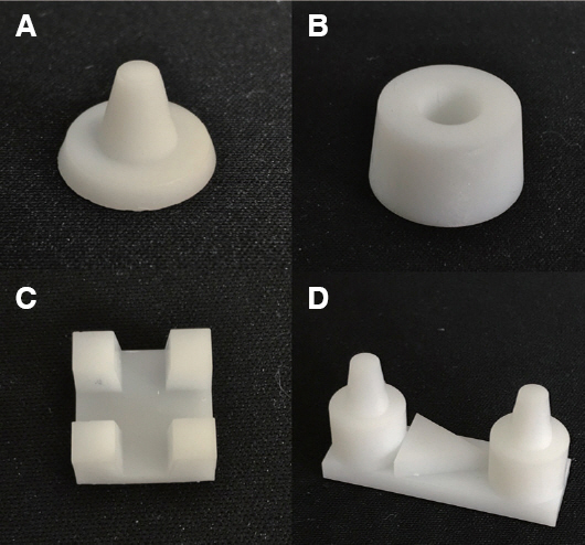
Fig. 1 The standard model image made with the 3D printer. (A) Crown model of ANSI/ADA 132, (B) inlay model of ANSI/ADA 132, (C) inlay model of ISO 12836 and (D) crown/bridge model of ISO 12836.
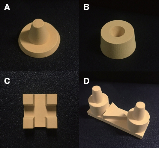
Fig. 2 The standard model image made with the improved stone. (A) Crown model of ANSI/ADA 132, (B) inlay model of ANSI/ADA 132, (C) inlay model of ISO 12836 and (D) crown/bridge model of ISO 12836.
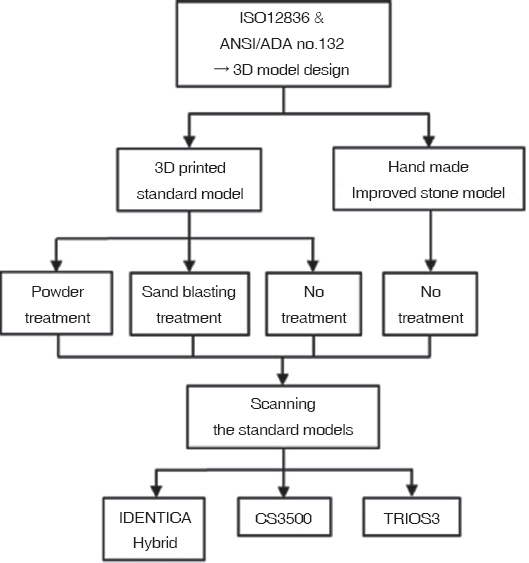
Fig. 3 A schematic diagram of the image acquisition of various scanners according to materials and surface conditions of the standard models.
Cited by 1 articles
-
Comparison of the accuracy of intraoral scanner by three-dimensional analysis in single and 3-unit bridge abutment model: In vitro study
Mei-Yang Huang, Keunbada Son, Wan-Sun Lee, Kyu-Bok Lee
J Korean Acad Prosthodont. 2019;57(2):102-109. doi: 10.4047/jkap.2019.57.2.102.
Reference
-
References
1. Miyazaki T, Hotta Y, Kunii J, Kuriyama S, Tamaki Y. A review of dental CAD/CAM:current status and future perspectives from 20 years of experience. Dent Mater J. 2009; 28:44–56. DOI: 10.4012/dmj.28.44. PMID: 19280967.2. Schleyer TK. Digital dentistry in the computer age. J Am Dent Assoc. 1999; 130:1713–20. DOI: 10.14219/jada.archive.1999.0127. PMID: 10599173.3. van Noort R. The future of dental devices is digital. Dent Mater. 2012; 28:3–12. DOI: 10.1016/j.dental.2011.10.014. PMID: 22119539.4. Davidowitz G, Kotick PG. The use of CAD/CAM in dentistry. Dent Clin North Am. 2011; 55:559–70. DOI: 10.1016/j.cden.2011.02.011. PMID: 21726690.5. Murad SM, Al-Mulla A. Accuracy of measurements made on digital and study models (A comparative study). MDJ. 2010; 7:71–82.6. Patzelt SB, Lamprinos C, Stampf S, Att W. The time efficiency of intraoral scanners:an in vitro comparative study. J Am Dent Assoc. 2014; 145:542–51. DOI: 10.14219/jada.2014.23. PMID: 24878708.7. Syrek A, Reich G, Ranftl D, Klein C, Cerny B, Brodesser J. Clinical evaluation of all-ceramic crowns fabricated from intraoral digital impressions based on the principle of active wave front sampling. J Dent. 2010; 38:553–9. DOI: 10.1016/j.jdent.2010.03.015. PMID: 20381576.8. Choi JH, Lim YJ, Lee WJ, Han JS, Lee SP. Review of recent developments for intra-oral scanners. J Dent Rehabil Appl Sci. 2015; 31:112–25. DOI: 10.14368/jdras.2015.31.2.112.9. Lee JJ, Park JY, Bae SY, Jeon JH, Kim JH, Kim WC. Evaluation of the Model Accuracy according to Three Types of Dental Scanner. J Dent Hyg Sci. 2015; 15:226–31. DOI: 10.17135/jdhs.2015.15.2.226.10. Schepke U, Meijer HJ, Kerdijk W, Cune MS. Digital versus analog complete-arch impressions for singleunit premolar implant crowns:Operating time and patient preference. J Prosthet Dent. 2015; 114:403–6. DOI: 10.1016/j.prosdent.2015.04.003. PMID: 26047800.11. Reddy MS, Mayfield-donahoo T, Vanderven FJ, Jeffcoat MK. A comparison of the diagnostic advantages of panoramic radiography and computed tomography scanning for placement of root form dental implants. Clin Oral Implants Res. 1994; 5:229–38. DOI: 10.1034/j.1600-0501.1994.050406.x. PMID: 7640337.12. Abdel-Azim T, Rogers K, Elathamna E, Zandinejad A, Metz M, Morton D. Comparison of the marginal fit of lithium disilicate crowns fabricated with CAD/CAM technology by using conventional impressions and two intraoral digital scanners. J Prosthet Dent. 2015; 114:554–9. DOI: 10.1016/j.prosdent.2015.04.001. PMID: 26100929.13. Ueda K, Beuer F, Stimmelmayr M, Erdelt K, Keul C, Güth JF. Fit of 4-unit FDPs from CoCr and zirconia after conventional and digital impressions. Clin Oral Investig. 2016; 20:283–9. DOI: 10.1007/s00784-015-1513-5. PMID: 26121970.14. Nedelcu RG, Persson AS. Scanning accuracy and precision in 4 intraoral scanners:an in vitro comparison based on 3-dimensional analysis. J Prosthet Dent. 2014; 112:1461–71. DOI: 10.1016/j.prosdent.2014.05.027. PMID: 25134995.15. Patzelt SB, Emmanouilidi A, Stampf S, Strub JR, Att W. Accuracy of full-arch scans using intraoral scanners. Clin Oral Investig. 2014; 18:1687–94. DOI: 10.1007/s00784-013-1132-y. PMID: 24240949.16. Patzelt SB, Vonau S, Stampf S, Att W. Assessing the feasibility and accuracy of digitizing edentulous jaws. J Am Dent Assoc. 2013; 144:914–20. DOI: 10.14219/jada.archive.2013.0209. PMID: 23904578.17. Papaspyridakos P, Gallucci GO, Chen CJ, Hanssen S, Naert I, Vandenberghe B. Digital versus conventional implant impressions for edentulous patients:accuracy outcomes. Clin Oral Implants Res. 2016; 27:465–72. DOI: 10.1111/clr.12567. PMID: 25682892.18. Lee GT, Kim JH, Kim WC, Kim JH. Three-dimensional evaluation on the repeatability and reproducibility of dental scanner-based digital models. J Korean Acad Dent Technol. 2012; 34:213–20. DOI: 10.14347/kadt.2012.34.3.213.19. Yuzbasioglu E, Kurt H, Turunc R, Bilir H. Comparison of digital and conventional impression techniques:evaluation of patients'perception, treatment comfort, effectiveness and clinical outcomes. BMC Oral Health. 2014; 14:10. DOI: 10.1186/1472-6831-14-10. PMID: 24479892. PMCID: PMC3913616.20. Kim JH, Kim KB. Evaluation of dimensional stability of digital dental model fabricated by impression scanning method. J Dent Hyg Sci. 2014; 14:15–21.21. Kim SH, Kim JH, Kim CK. Reliability and accuracy of digital impression obtained from CS-3500 intraoral scanner. J Dent Hyg Sci. 2015; 15:673–8. DOI: 10.17135/jdhs.2015.15.5.673.22. ISO 12836:2015 Dentistry - Digitizing devices for CAD/CAM systems for indirect dental restorations - Test methods for assessing accuracy.23. ANSI/ADA. Standard No. 132, Scanning accuracy of dental chairside and laboratory CAD/CAM. ADA 132-2015.
- Full Text Links
-
- Actions
-
Cited
- CITED
-
- Close
- Share
-
- Similar articles
-
- Erratum: Comparative study on quality of scanned images from varying materials and surface conditions of standardized model for dental scanner evaluation
- Influence of orthodontic bracket block-out materials on superimposition errors when substituting scanned dental imaging data onto computed tomography images
- Comparative analysis on reproducibility among 5 intraoral scanners: sectional analysis according to restoration type and preparation outline form
- Evaluation of the reproducibility of various abutments using a blue light model scanner
- Clinical application of an intraoral scanner for serial evaluation of orthodontic tooth movement: A preliminary study
