Surgical Treatment of Macrodystrophia Lipomatosa Involving Superficial Peroneal Nerve in the Foot and Ankle
- Affiliations
-
- 1Department of Orthopaedic Surgery, Samsung Changwon Hospital, Changwon, Korea. orthoshin@gmail.com
- KMID: 2421352
- DOI: http://doi.org/10.4055/jkoa.2017.52.6.566
Abstract
- Macrodystrophia lipomatosa is a congenital disease characterized by gradual proliferation in the mesenchymal cell, such as fibroadipose tissue. Pathologically, fatty tissue is deposited in the nerve sheath, periosteum, bone marrow, and subcutaneous tissue, contributing to the macrodactyly of the foot. To date, there has not been any report on macrodystrophia lipomatosa of the superficial peroneal nerve in the Korean orthopedic literature. Conservative approach, such as decompression or debulking surgery, is recommended due to neurogenic dysfunction. However, we report a 43-year-old male with macrodystrophia lipomatosa involving the superficial peroneal nerve of the right foot and ankle, who underwent a second toe ray amputation as well as soft tissue and nerve resection.
MeSH Terms
Figure
-
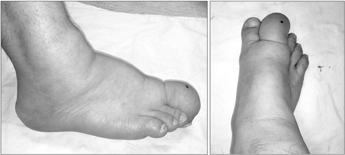
Figure 1 Photographs showing cordlike mass on the right foot and ankle with second toe macrodactyly.
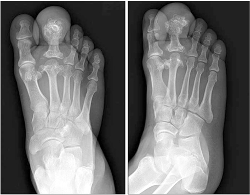
Figure 2 Radiographs showing interphalangeal and metatarsophalangeal joint arthritis with hypertrophied phalanx and multiple bony spur.

Figure 3 Coronal magnetic resonance imaging of the forefoot showing an enlarged superficial peroneal nerve (white arrows). (A) Nerve fascicles of the superficial peroneal nerve are surrounded by fat tissue with high signal intensity within nerve sheath on T1-weighted imaging. T2-weighted image showing fat tissue with intermediate signal intensity (B) and low signal intensity on T2 fat suppressed image (C).
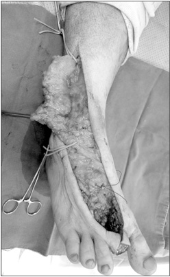
Figure 4 Intraoperative photograph showing medial dorsal cutaneous branch of the superficial peroneal nerve, which was hypertrophied with fatty tissue.
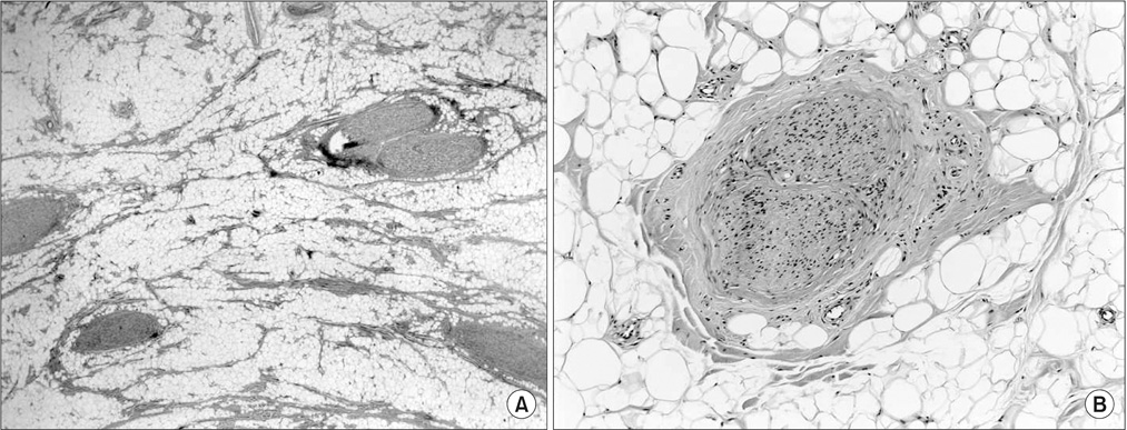
Figure 5 (A) Infiltration of the epineurium and perineurium by adipose and fibrous tissue, causing an enlargement of the nerve (H&E, ×12.5). (B) Concentric perineurial fibrous tissue and pseudo-onion bulb formation (H&E, ×100).
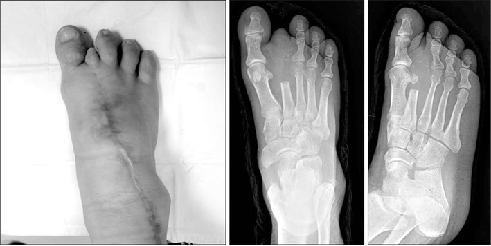
Figure 6 The last follow-up photograph and radiographs of the foot showing no evidence of recurrence.
Reference
-
1. Moran V, Butler F, Colville J. X-ray diagnosis of macrodystrophia lipomatosa. Br J Radiol. 1984; 57:523–525.2. Khan RA, Wahab S, Ahmad I, Chana RS. Macrodystrophia lipomatosa: four case reports. Ital J Pediatr. 2010; 36:69.3. Ho CA, Herring JA, Ezaki M. Long-term follow-up of progressive macrodystrophia lipomatosa. A report of two cases. J Bone Joint Surg Am. 2007; 89:1097–1102.4. Fransen BL, Broeders MG, van Oosterom FJ, Gilhuijs ND, Burger BJ. Operative resection is a viable treatment of macrodactyly of the foot caused by lipofibromatous hamartoma: a case study with 5 year follow-up. Foot Ankle Surg. 2014; 20:e47–e50.5. Goldman AB, Kaye JJ. Macrodystrophia lipomatosa: radiographic diagnosis. AJR Am J Roentgenol. 1977; 128:101–105.6. Dhinsa BS, Lidder S, Abbasian A. Atypical presentation of fibrolipomatous hamartoma of superficial peroneal nerve. J Foot Ankle Surg. 2016; 55:1067–1068.7. Çelebi F, Karagulle K, Oner AY. Macrodystrophia lipomatosa of the foot: a case report. Oncol Lett. 2015; 10:951–953.8. Lee MJ, Kim DR. Recurred macrodystrophia lipomatosa of the foot: a case report. J Korean Foot Ankle Soc. 2016; 20:32–35.9. Razzaghi A, Anastakis DJ. Lipofibromatous hamartoma: review of early diagnosis and treatment. Can J Surg. 2005; 48:394–399.10. Brodwater BK, Major NM, Goldner RD, Layfield LJ. Macrodystrophia lipomatosa with associated fibrolipomatous hamartoma of the median nerve. Pediatr Surg Int. 2000; 16:216–218.
- Full Text Links
-
- Actions
-
Cited
- CITED
-
- Close
- Share
-
- Similar articles
-
- Macrodystrophia Lipomatosa of the Foot (A Case Report)
- Recurred Macrodystrophia Lipomatosa of the Foot: A Case Report
- Recurred Intraneural Ganglion on Superficial Peroneal Nerve (A Case Report)
- Entrapment of Superficial Peroneal Nerve (A Case Report)
- Superficial Peroneal Nerve Entrapment Syndrome (A Case Report)
