Feasibility of Quadruple Arterial Phase of Motion Insensitive Radial Volumetric Imaging Breath-Hold Examination with k-Space Weighted Image Contrast in the Detection of Hepatocellular Carcinoma in Patients with Chronic Liver Disease
- Affiliations
-
- 1Department of Radiology, Jeju National University Hospital, Jeju National University School of Medicine, Jeju, Korea. 671228kbs@naver.com
- KMID: 2421220
- DOI: http://doi.org/10.3348/jksr.2018.79.4.181
Abstract
- PURPOSE
To evaluate the detection performance of hepatocellular carcinoma and image quality in patients with chronic liver disease with quadruple arterial MR imaging using radial volumetric imaging breath-hold examination (VIBE) with k-space weighted image contrast (KWIC).
MATERIALS AND METHODS
Forty-four patients underwent liver MR examinations with quadruple arterial imaging using radial VIBE-KWIC sequence (full-frame and four sub-frame images). Diagnostic performance was evaluated with receiver operating characteristics (ROC) for detection of hepatocellular carcinoma. The image quality and severity of artifact were scored by using the five-point scale.
RESULTS
The area under the ROC curve (Az) value of Hepatocelluar Carcinoma (HCC) detectability was the highest on third sub-frame images, followed by full-frame images. The Az values of third sub-frame and full-frame about the detection of HCC were statistically significantly different from the Az value of first sub-frame images. The full-frame and four sub-frame images showed acceptable image quality and low degree artifact with rating of higher than grade 3.
CONCLUSION
Quadruple arterial MRI using radial VIBE-KWIC is a feasible method for detecting hepatocellular carcinoma in patients with chronic liver disease without deterioration of image quality. The third sub-frame and full-frame image are superior to other sub-frame images in detecting hepatocellular carcinoma.
MeSH Terms
Figure
-
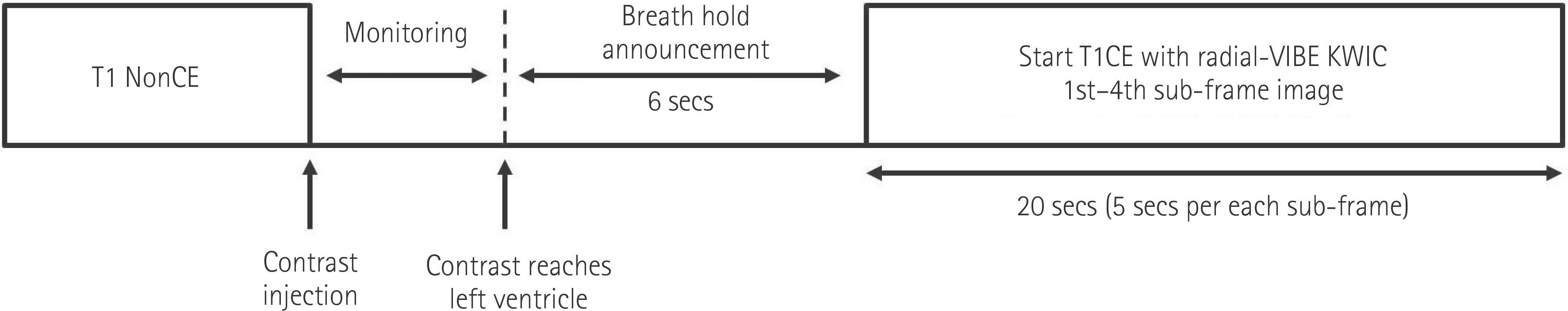
Fig. 1 Schematic timing diagram for T1-weighted quadruple arterial MR imaging using radial VIBE-KWIC. KWIC = k-space weighted image contrast, T1CE = contrast-enhanced T1-weighted image, VIBE = volumetric imaging breath-hold examination

Fig. 2 Principles of the radial acquisition with KWIC reconstruction technique. A. Schematic diagram of the four-interleaf angle-bisection reordering acquisition shows simple example composed of eight projection views. For making full-frame image, k-space data are serially obtained radially projection views which are grouped into four interleaved subsets. And data from all subsets are combined for the reconstruction of full-frame images. B. For the reconstruction of sub-frame images, KWIC divides the k-space into three regions based on the circular Nyquist radius and fills the spokes. For example, spokes obtained first among all acquired radial spokes are filled in k-space. The next spokes are filled without the center. The last obtained spokes are used to fill only the outermost part of the k-space so that it has little effect on the resolution. Therefore four sub-frame KWIC images have different k-space cores and the surrounding k-space is similar. KWIC = k-space weighted image contrast

Fig. 3 MR images obtained in a 64-year-old man with a hepatocellular carcinoma. Contrast-enhanced 3D Radial k-space weighted image contrast, volumetric imaging breath-hold examination during hepatic arterial dominant phase imaging comprised one full-frame and four sub-frame images (A-E). The full-frame and sub-frame images show a focal enhancing lesion (arrows) in right hepatic lobe. Third sub-frame image (D) shows tumor with greater conspicuity and best arterial enhancement than other arterial images (A-C, E). Much better image quality and lesser artifacts were seen on full-frame image. The arterial enhancing lesion was pathologically proved to be a hepatocellular carcinoma after right anterior sectionectomy.

Fig. 4 MR and CT images obtained in a 63-year-old man with a hepatocellular carcinoma. Full-frame and four sub-frame images using contrast-enhanced 3D Radial k-space weighted image contrast, volumetric imaging breath-hold examination during multiple hepatic arterial dominant phases at 5-second temporal resolution (A-E), show gradually increasing enhancing lesion (arrows) in left hepatic lobe. Third sub-frame image (D) depicts an enhancing tumor (arrow) more clearly with best arterial enhancement than other arterial images (A-C, E). Three months follow-up CT (F) after transcatheter arterial chemoembolization demonstrates accumulation of iodized oil (arrow) in a hepatocellular carcinoma in left hepatic lobe. TACE = transcatheter arterial chemoembolizatio
Reference
-
1.Hawighorst H., Schoenberg SO., Knopp MV., Essig M., Miltner P., van Kaick G. Hepatic lesions: morphologic and functional characterization with multiphase breath-hold 3D gadolinium-enhanced MR angiography—initial results. Radiology. 1999. 210:89–96.
Article2.Elsayes KM., Narra VR., Yin Y., Mukundan G., Lammle M., Brown JJ. Focal hepatic lesions: diagnostic value of enhancement pattern approach with contrast-enhanced 3D gradient-echo MR imaging. Radiographics. 2005. 25:1299–1320.
Article3.Kim BK., Kim MJ., Park BJ., Sung DJ., Cho SB. [Triple arterial phase hepatic MRI using four dimensional T1-weighted high resolutions imaging with volume excitation keyhole techniques: feasibility and initial clinical experience in focal liver lesions]. J Korean Soc Radiol. 2013. 69:223–234.
Article4.Hong HS., Kim HS., Kim MJ., De Becker J., Mitchell DG., Kanematsu M. Single breath-hold multiarterial dynamic MRI of the liver at 3T using a 3D fat-suppressed keyhole technique. J Magn Reson Imaging. 2008. 28:396–402.
Article5.Kanematsu M., Semelka RC., Matsuo M., Kondo H., Enya M., Goshima S, et al. Gadolinium-enhanced MR imaging of the liver: optimizing imaging delay for hepatic arterial and portal venous phases—a prospective randomized study in patients with chronic liver damage. Radiology. 2002. 225:407–415.
Article6.Goshima S., Kanematsu M., Kondo H., Yokoyama R., Miyoshi T., Nishibori H, et al. MDCT of the liver and hypervascular hepatocellular carcinomas: optimizing scan delays for bolus-tracking techniques of hepatic arterial and portal venous phases. AJR Am J Roentgenol. 2006. 187:W25–W32.
Article7.Kim KW., Lee JM., Jeon YS., Kang SE., Baek JH., Han JK, et al. Free-breathing dynamic contrast-enhanced MRI of the abdomen and chest using a radial gradient echo sequence with K-space weighted image contrast (KWIC). Eur Radiol. 2013. 23:1352–1360.
Article8.Fujinaga Y., Ohya A., Tokoro H., Yamada A., Ueda K., Ueda H, et al. Radial volumetric imaging breath-hold examination (VIBE) with k-space weighted image contrast (KWIC) for dynamic gadoxetic acid (Gd-EOB-DTPA)-enhanced MRI of the liver: advantages over Cartesian VIBE in the arterial phase. Eur Radiol. 2014. 24:1290–1299.
Article9.Brodsky EK., Bultman EM., Johnson KM., Horng DE., Schelman WR., Block WF, et al. High-spatial and high-temporal resolution dynamic contrast-enhanced perfusion imaging of the liver with time-resolved three-dimensional radial MRI. Magn Reson Med. 2014. 71:934–941.
Article10.Zech CJ., Vos B., Nordell A., Urich M., Blomqvist L., Breuer J, et al. Vascular enhancement in early dynamic liver MR imaging in an animal model: comparison of two injection regimen and two different doses Gd-EOB-DTPA (gadoxetic acid) with standard Gd-DTPA. Invest Radiol. 2009. 44:305–310.
Article11.Park YS., Lee CH., Yoo JL., Kim IS., Kiefer B., Woo ST, et al. Hepatic arterial phase in gadoxetic acid-enhanced liver magnetic resonance imaging: analysis of respiratory patterns and their effect on image quality. Invest Radiol. 2016. 51:127–133.12.Hope TA., Saranathan M., Petkovska I., Hargreaves BA., Her-fkens RJ., Vasanawala SS. Improvement of gadoxetate arterial phase capture with a high spatio-temporal resolution multiphase three-dimensional SPGR-Dixon sequence. J Magn Reson Imaging. 2013. 38:938–945.
Article13.Beck GM., De Becker J., Jones AC., von Falkenhausen M., Wil-linek WA., Gieseke J. Contrast-enhanced timing robust acquisition order with a preparation of the longitudinal signal component (CENTRA plus) for 3D contrast-enhanced abdominal imaging. J Magn Reson Imaging. 2008. 27:1461–1467.
Article14.Hadizadeh DR., Gieseke J., Beck G., Geerts L., Kukuk GM., Boström A, et al. View-sharing in keyhole imaging: partially compressed central k-space acquisition in time-resolved MRA at 3.0 T. Eur J Radiol. 2011. 80:400–406.15.Agrawal MD., Spincemaille P., Mennitt KW., Xu B., Wang Y., Dutruel SP, et al. Improved hepatic arterial phase MRI with 3-second temporal resolution. J Magn Reson Imaging. 2013. 37:1129–1136.
Article16.Kim BS., Lee KR., Goh MJ. New imaging strategies using a motion-resistant liver sequence in uncooperative patients. Biomed Res Int. 2014. 2014:142658.
Article17.Budjan J., Riffel P., Ong MM., Schoenberg SO., Attenberger UI., Hausmann D. Rapid Cartesian versus radial acquisition: comparison of two sequences for hepatobiliary phase MRI at 3 tesla in patients with impaired breath-hold capabilities. BMC Med Imaging. 2017. 17:32.
Article18.Yu MH., Lee JM., Yoon JH., Kiefer B., Han JK., Choi BI. Clinical application of controlled aliasing in parallel imaging results in a higher acceleration (CAIPIRINHA)-volumetric interpolated breathhold (VIBE) sequence for gadoxetic acid-enhanced liver MR imaging. J Magn Reson Imaging. 2013. 38:1020–1026.
Article19.Li H., Xiao Y., Wang S., Li Y., Zhong X., Situ W, et al. TWIST-VIBE five-arterial-phase technology decreases transient severe motion after bolus injection of Gd-EOB-DTPA. Clin Radiol. 2017. 72:800. .e1-800.e6.
Article20.Theilmann RJ., Gmitro AF., Altbach MI., Trouard TP. View-ordering in radial fast spin-echo imaging. Magn Reson Med. 2004. 51:768–774.
Article21.Spuentrup E., Katoh M., Buecker A., Manning WJ., Schaeffter T., Nguyen TH, et al. Free-breathing 3D steady-state free precession coronary MR angiography with radial k-space sampling: comparison with cartesian k-space sampling and cartesian gradient-echo coronary MR angiography—pilot study. Radiology. 2004. 231:581–586.
Article22.Clarke SE., Saranathan M., Rettmann DW., Hargreaves BA., Vasanawala SS. High resolution multiarterial phase MRI improves lesion contrast in chronic liver disease. Clin Invest Med. 2015. 38:E90–E99.
Article23.Pietryga JA., Burke LM., Marin D., Jaffe TA., Bashir MR. Respiratory motion artifact affecting hepatic arterial phase imaging with gadoxetate disodium: examination recovery with a multiple arterial phase acquisition. Radiology. 2014. 271:426–434.
Article
- Full Text Links
-
- Actions
-
Cited
- CITED
-
- Close
- Share
-
- Similar articles
-
- Advanced Methods in Dynamic Contrast Enhanced Arterial Phase Imaging of the Liver
- Triple Arterial Phase Hepatic MRI Using Four Dimensional T1-Weighted High Resolutions Imaging with Volume Excitation Keyhole Techniques: Feasibility and Initial Clinical Experience in Focal Liver Lesions
- Detection of Hepatocelluar Carcinoma on Triple-Phase Images of Liver Using Multi-Detector Row Helical CT
- Comparison of In-Phase and Opposed-Phase FMPSPGR Images in Breath-hold T1-weighted MR Imaging of Liver
- Comparison of Non-Breath-Hold T2-weighted Turbo Spin-Echo and Three Breath-Hold T2-weighted MR Images for Detection of Focal Hepatic Lesion
