CT-Guided Percutaneous Transthoracic Needle Biopsy Using the Additional Laser Guidance System by a Pulmonologist with 2 Years of Experience in CT-Guided Percutaneous Transthoracic Needle Biopsy
- Affiliations
-
- 1Department of Radiology, Daejeon Health Institute of Technology, Daejeon, Korea.
- 2Division of Pulmonary and Critical Care Medicine, Department of Internal Medicine, Chungnam National University Hospital, Daejeon, Korea. rahm3s@gmail.com
- KMID: 2420567
- DOI: http://doi.org/10.4046/trd.2017.0123
Abstract
- BACKGROUND
We developed an additional laser guidance system to improve the efficacy and safety of conventional computed tomography (CT)-guided percutaneous transthoracic needle biopsy (PTNB), and we conducted this study to evaluate the efficacy and safety of our system.
METHODS
We retrospectively analyzed the medical records of 244 patients who underwent CT-guided PTNB using our additional laser guidance system from July 1, 2015, to January 20, 2016.
RESULTS
There were nine false-negative results among the 238 total cases. The sensitivity, specificity, positive predictive value, negative predictive value, and diagnostic accuracy of our system for diagnosing malignancy were 94.4% (152/161), 100% (77/77), 100% (152/152), 89.5% (77/86), and 96.2% (229/238), respectively. The results of univariate analysis showed that the risk factors for a false-negative result were male sex (p=0.029), a final diagnosis of malignancy (p=0.033), a lesion in the lower lobe (p=0.035), shorter distance from the skin to the target lesion (p=0.003), and shorter distance from the pleura to the target lesion (p=0.006). The overall complication rate was 30.5% (74/243). Pneumothorax, hemoptysis, and hemothorax occurred in 21.8% (53/243), 9.1% (22/243), and 1.6% (4/243) of cases, respectively.
CONCLUSION
The additional laser guidance system might be a highly economical and efficient method to improve the diagnostic efficacy and safety of conventional CT-guided PTNB even if performed by inexperienced pulmonologists.
MeSH Terms
Figure
-
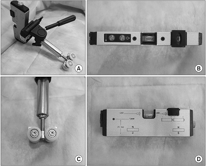
Figure 1 Laser level (HG-909A; X-CLOVE) and a wheel bracket. (A) Laser level, adhered to a wheel bracket. (B, D) Overhead and side view of laser level. (C) Wheel part of a bracket.
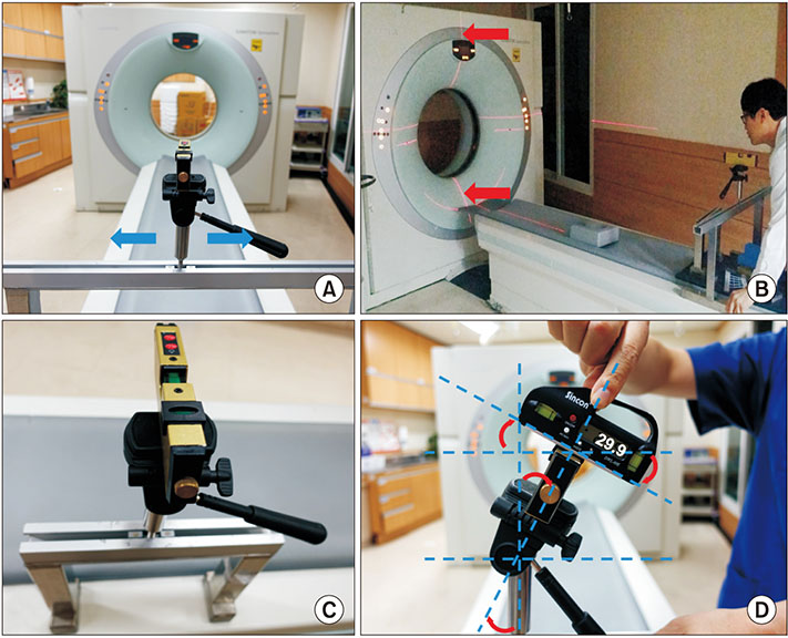
Figure 2 (A) The laser level, adhered to a wheel bracket, mounted on a stainless steel frame and positioned on the opposite side of the computed tomography (CT) gantry. Horizontal movement is possible (blue arrows). (B) Both vertical laser beam lines, which came from the laser level and CT gantry, had been aligned, such that they looked like a line (red arrows). (C) The second laser level was placed on the side of the patient opposite to the operator, perpendicular to the other laser. (D) In the case of an oblique approach, a digital level (DWL-80E, Sincon) was used to tilt the laser level. The digital level was 29.9° (red round arrows).
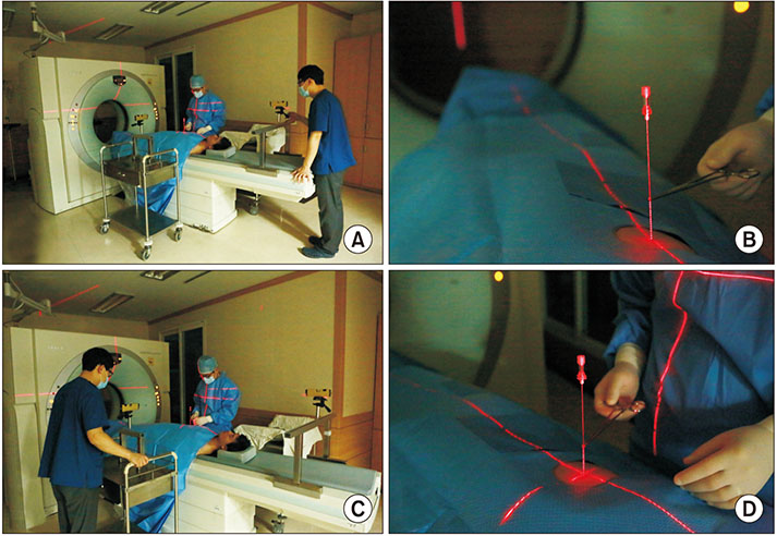
Figure 3 Adjustment of the laser lines. (A, B) The laser line originating from the head end was located at the entry point on the skin and ran along the coaxial needle. (C, D) The intersection of the two laser lines originating from the head end and the side opposite to the operator were matched at the entry point on the skin and ran long the coaxial needle.
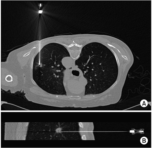
Figure 4 A 76-year-old woman who was diagnosed with non-small cell lung cancer (adenocarcinoma) by percutaneous transthoracic needle biopsy. (A) Axial view. (B) Sagittal view.
Reference
-
1. DiBardino DM, Yarmus LB, Semaan RW. Transthoracic needle biopsy of the lung. J Thorac Dis. 2015; 7:Suppl 4. S304–S316.2. Jin KN, Park CM, Goo JM, Lee HJ, Lee Y, Kim JI, et al. Initial experience of percutaneous transthoracic needle biopsy of lung nodules using C-arm cone-beam CT systems. Eur Radiol. 2010; 20:2108–2115.
Article3. Lee SM, Park CM, Lee KH, Bahn YE, Kim JI, Goo JM. C-arm cone-beam CT-guided percutaneous transthoracic needle biopsy of lung nodules: clinical experience in 1108 patients. Radiology. 2014; 271:291–300.
Article4. Yan GW, Bhetuwal A, Yan GW, Sun QQ, Niu XK, Zhou Y, et al. A systematic review and meta-analysis of C-arm cone-beam CT-guided percutaneous transthoracic needle biopsy of lung nodules. Pol J Radiol. 2017; 82:152–160.
Article5. Zhuang YP, Wang HY, Zhang J, Feng Y, Zhang L. Diagnostic accuracy and safety of CT-guided fine needle aspiration biopsy in cavitary pulmonary lesions. Eur J Radiol. 2013; 82:182–186.
Article6. Li Y, Du Y, Yang HF, Yu JH, Xu XX. CT-guided percutaneous core needle biopsy for small (≤20 mm) pulmonary lesions. Clin Radiol. 2013; 68:e43–e48.7. Loh SE, Wu DD, Venkatesh SK, Ong CK, Liu E, Seto KY, et al. CT-guided thoracic biopsy: evaluating diagnostic yield and complications. Ann Acad Med Singapore. 2013; 42:285–290.8. Takeshita J, Masago K, Kato R, Hata A, Kaji R, Fujita S, et al. CT-guided fine-needle aspiration and core needle biopsies of pulmonary lesions: a single-center experience with 750 biopsies in Japan. AJR Am J Roentgenol. 2015; 204:29–34.
Article9. Yang W, Sun W, Li Q, Yao Y, Lv T, Zeng J, et al. Diagnostic accuracy of CT-guided transthoracic needle biopsy for solitary pulmonary nodules. PLoS One. 2015; 10:e0131373.
Article10. Yaffe D, Koslow M, Haskiya H, Shitrit D. A novel technique for CT-guided transthoracic biopsy of lung lesions: improved biopsy accuracy and safety. Eur Radiol. 2015; 25:3354–3360.
Article11. Choi SH, Chae EJ, Kim JE, Kim EY, Oh SY, Hwang HJ, et al. Percutaneous CT-guided aspiration and core biopsy of pulmonary nodules smaller than 1 cm: analysis of outcomes of 305 procedures from a tertiary referral center. AJR Am J Roentgenol. 2013; 201:964–970.
Article12. Choo JY, Park CM, Lee NK, Lee SM, Lee HJ, Goo JM. Percutaneous transthoracic needle biopsy of small (≤1 cm) lung nodules under C-arm cone-beam CT virtual navigation guidance. Eur Radiol. 2013; 23:712–719.13. Hwang HS, Chung MJ, Lee JW, Shin SW, Lee KS. C-arm conebeam CT-guided percutaneous transthoracic lung biopsy: usefulness in evaluation of small pulmonary nodules. AJR Am J Roentgenol. 2010; 195:W400–W407.
Article14. Larke FJ, Kruger RL, Cagnon CH, Flynn MJ, McNitt-Gray MM, Wu X, et al. Estimated radiation dose associated with low-dose chest CT of average-size participants in the National Lung Screening Trial. AJR Am J Roentgenol. 2011; 197:1165–1169.
Article15. Hiraki T, Mimura H, Gobara H, Iguchi T, Fujiwara H, Sakurai J, et al. CT fluoroscopy-guided biopsy of 1,000 pulmonary lesions performed with 20-gauge coaxial cutting needles: diagnostic yield and risk factors for diagnostic failure. Chest. 2009; 136:1612–1617.16. Chami HA, Faraj W, Yehia ZA, Badour SA, Sawan P, Rebeiz K, et al. Predictors of pneumothorax after CT-guided transthoracic needle lung biopsy: the role of quantitative CT. Clin Radiol. 2015; 70:1382–1387.
Article17. Boskovic T, Stanic J, Pena-Karan S, Zarogoulidis P, Drevelegas K, Katsikogiannis N, et al. Pneumothorax after transthoracic needle biopsy of lung lesions under CT guidance. J Thorac Dis. 2014; 6:Suppl 1. S99–S107.18. van Haren-Willems J, Heijdra Y. Increasing evidence for gender differences in chronic obstructive pulmonary disease. Womens Health (Lond). 2010; 6:595–600.
Article19. Yeow KM, Tsay PK, Cheung YC, Lui KW, Pan KT, Chou AS. Factors affecting diagnostic accuracy of CT-guided coaxial cutting needle lung biopsy: retrospective analysis of 631 procedures. J Vasc Interv Radiol. 2003; 14:581–588.
Article20. Ko JP, Shepard JO, Drucker EA, Aquino SL, Sharma A, Sabloff B, et al. Factors influencing pneumothorax rate at lung biopsy: are dwell time and angle of pleural puncture contributing factors? Radiology. 2001; 218:491–496.
Article21. Khan MF, Straub R, Moghaddam SR, Maataoui A, Gurung J, Wagner TO, et al. Variables affecting the risk of pneumothorax and intrapulmonal hemorrhage in CT-guided transthoracic biopsy. Eur Radiol. 2008; 18:1356–1363.
Article22. Rotolo N, Floridi C, Imperatori A, Fontana F, Ierardi AM, Mangini M, et al. Comparison of cone-beam CT-guided and CT fluoroscopy-guided transthoracic needle biopsy of lung nodules. Eur Radiol. 2016; 26:381–389.
Article23. Adiga S, Athreya S. Safety, efficacy, and feasibility of an ultra-low dose radiation protocol for CT-guided percutaneous needle biopsy of pulmonary lesions: initial experience. Clin Radiol. 2014; 69:709–714.
Article24. Lechuga L, Weidlich GA. Cone beam CT vs. fan beam CT: a comparison of image quality and dose delivered between two differing CT imaging modalities. Cureus. 2016; 8:e778.
Article25. Schegerer AA, Lechel U, Ritter M, Weisser G, Fink C, Brix G. Dose and image quality of cone-beam computed tomography as compared with conventional multislice computed tomography in abdominal imaging. Invest Radiol. 2014; 49:675–684.
Article26. Ravenel JG, Scalzetti EM, Huda W, Garrisi W. Radiation exposure and image quality in chest CT examinations. AJR Am J Roentgenol. 2001; 177:279–284.
Article27. Hirota S, Nakao N, Yamamoto S, Kobayashi K, Maeda H, Ishikura R, et al. Cone-beam CT with flat-panel-detector digital angiography system: early experience in abdominal interventional procedures. Cardiovasc Intervent Radiol. 2006; 29:1034–1038.
Article
- Full Text Links
-
- Actions
-
Cited
- CITED
-
- Close
- Share
-
- Similar articles
-
- Hematoma-Filled Pneumatocele after CT-Guided Percutaneous Transthoracic Needle Lung Biopsy: Two Case Reports
- Painful Percutaneous Transthoracic Needle Biopsy of Schwannoma: A Case Report
- Air Embolization after Computed Tomography-Guided Percutaneous Transthoracic Needle Biopsy
- Analysis of the result and merit of computed tomography guided percutaneous needle aspiration biopsy of focal lung lesion
- Transthoracic Needle Biopsy of Thoracic Lesions
