Yonsei Med J.
2018 Oct;59(8):937-944. 10.3349/ymj.2018.59.8.937.
Clinical Implications of Moderate Coronary Stenosis on Coronary Computed Tomography Angiography in Patients with Stable Angina
- Affiliations
-
- 1Division of Cardiology, Severance Cardiovascular Hospital, Yonsei University College of Medicine, Seoul, Korea. mkhong61@yuhs.ac
- 2Cardiovascular Research Institute, Yonsei University College of Medicine, Seoul, Korea.
- 3Division of Cardiovascular Radiology, Severance Cardiovascular Hospital, Yonsei University College of Medicine, Seoul, Korea.
- 4Department of Radiology, Yonsei University College of Medicine, Seoul, Korea.
- KMID: 2419727
- DOI: http://doi.org/10.3349/ymj.2018.59.8.937
Abstract
- PURPOSE
The present study investigated the diagnostic accuracy and clinical implications of moderate stenosis (50-69%, Coronary Artery Disease Reporting and Data System, grade 3) on coronary computed tomography angiography (CCTA), compared with invasive coronary angiography (ICA).
MATERIALS AND METHODS
Two hundred and seventy-six patients who underwent ICA due to moderate stenosis alone on CCTA were selected from our prospective registry cohort.
RESULTS
Diagnostic concordance between CCTA and ICA was found in only 50 (18%) patients. Among the 396 vessels and 508 segments with moderate stenosis, diagnostic concordance was found in 132 vessels (33%) and 127 segments (25%). Segments with calcified plaque had lower diagnostic concordance than those with mixed or non-calcified plaque (22% vs. 28% vs. 27%, respectively, p=0.001). While calcified plaque burden did not have an influence on severe stenosis (≥70%) on ICA, higher burden of non-calcified plaque was correlated with a greater incidence of ICA-based severe stenosis, which was more frequent in patients with ≥3 segments of non-calcified plaque (75%) than those without non-calcified plaque (22%, p < 0.001). Typical angina and mixed or non-calcified plaque were correlated with a higher incidence of under-diagnosis, while the use of next-generation computed tomography scanners reduced the incidence of under-diagnosis. Increased body weight, left circumflex artery involvement, and calcified plaque were independent factors that increased the risk of over-diagnosis of CCTA.
CONCLUSION
The diagnosis of moderate stenosis by CCTA may be limited in estimating the exact degree of ICA-based anatomical stenosis. Unlike calcific burden, non-calcific burden was positively correlated with the presence of severe stenosis on ICA.
MeSH Terms
Figure
-
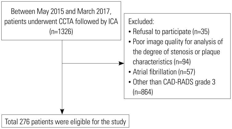
Fig. 1 Study flow. CAD-RADS, Coronary Artery Disease Reporting and Data System; CCTA, coronary computed tomography angiography; ICA, invasive coronary angiography.

Fig. 2 Diagnostic correlation between CCTA and ICA per patient analysis. (A) Diagnostic accuracy. (B) Degree of maximal stenosis. CCTA, coronary computed tomography angiography; ICA, invasive coronary angiography; VD, vessel disease.
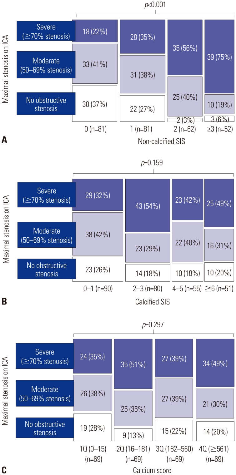
Fig. 3 Presence of any significant stenosis in patients according to plaque burden. Patients were categorized by quartiles of non-calcified (A) and calcified (B) SIS, and calcium score (C). CT, computed tomography; SIS, segment involvement score; ICA, invasive coronary angiography.
Reference
-
1. Miller JM, Rochitte CE, Dewey M, Arbab-Zadeh A, Niinuma H, Gottlieb I, et al. Diagnostic performance of coronary angiography by 64-row CT. N Engl J Med. 2008; 359:2324–2336. PMID: 19038879.
Article2. Litt HI, Gatsonis C, Snyder B, Singh H, Miller CD, Entrikin DW, et al. CT angiography for safe discharge of patients with possible acute coronary syndromes. N Engl J Med. 2012; 366:1393–1403. PMID: 22449295.
Article3. SCOT-HEART investigators. CT coronary angiography in patients with suspected angina due to coronary heart disease (SCOTHEART): an open-label, parallel-group, multicentre trial. Lancet. 2015; 385:2383–2391. PMID: 25788230.4. Cury RC, Abbara S, Achenbach S, Agatston A, Berman DS, Budoff MJ, et al. CAD-RADS(TM) Coronary Artery Disease - Reporting and Data System. An expert consensus document of the Society of Cardiovascular Computed Tomography (SCCT), the American College of Radiology (ACR) and the North American Society for Cardiovascular Imaging (NASCI). Endorsed by the American College of Cardiology. J Cardiovasc Comput Tomogr. 2016; 10:269–281. PMID: 27318587.
Article5. Pryor DB, Harrell FE Jr, Lee KL, Califf RM, Rosati RA. Estimating the likelihood of significant coronary artery disease. Am J Med. 1983; 75:771–780. PMID: 6638047.
Article6. Taylor AJ, Cerqueira M, Hodgson JM, Mark D, Min J, O'Gara P, et al. ACCF/SCCT/ACR/AHA/ASE/ASNC/NASCI/SCAI/SCMR 2010 appropriate use criteria for cardiac computed tomography. A report of the American College of Cardiology Foundation Appropriate Use Criteria Task Force, the Society of Cardiovascular Computed Tomography, the American College of Radiology, the American Heart Association, the American Society of Echocardiography, the American Society of Nuclear Cardiology, the North American Society for Cardiovascular Imaging, the Society for Cardiovascular Angiography and Interventions, and the Society for Cardiovascular Magnetic Resonance. J Am Coll Cardiol. 2010; 56:1864–1894. PMID: 21087721.7. Austen WG, Edwards JE, Frye RL, Gensini GG, Gott VL, Griffith LS, et al. A reporting system on patients evaluated for coronary artery disease. Report of the Ad Hoc Committee for Grading of Coronary Artery Disease, Council on Cardiovascular Surgery, American Heart Association. Circulation. 1975; 51(4 Suppl):5–40. PMID: 1116248.
Article8. Hong SJ, Her AY, Suh Y, Won H, Cho DK, Cho YH, et al. Coronary computed tomographic angiography does not accurately predict the need of coronary revascularization in patients with stable angina. Yonsei Med J. 2016; 57:1079–1086. PMID: 27401637.
Article9. Achenbach S, Raggi P. Imaging of coronary atherosclerosis by computed tomography. Eur Heart J. 2010; 31:1442–1448. PMID: 20484566.
Article10. Groothuis JG, Beek AM, Meijerink MR, Brinckman SL, Heymans MW, van Kuijk C, et al. Positive predictive value of computed tomography coronary angiography in clinical practice. Int J Cardiol. 2012; 156:315–319. PMID: 21195491.
Article11. Dey D, Lee CJ, Ohba M, Gutstein A, Slomka PJ, Cheng V, et al. Image quality and artifacts in coronary CT angiography with dual-source CT: initial clinical experience. J Cardiovasc Comput Tomogr. 2008; 2:105–114. PMID: 19083930.
Article12. Øvrehus KA, Marwan M, Bøtker HE, Achenbach S, Nørgaard BL. Reproducibility of coronary plaque detection and characterization using low radiation dose coronary computed tomographic angiography in patients with intermediate likelihood of coronary artery disease (ReSCAN study). Int J Cardiovasc Imaging. 2012; 28:889–899. PMID: 21626043.
Article13. McHugh ML. Interrater reliability: the kappa statistic. Biochem Med (Zagreb). 2012; 22:276–282. PMID: 23092060.
Article14. Shrout PE, Fleiss JL. Intraclass correlations: uses in assessing rater reliability. Psychol Bull. 1979; 86:420–428. PMID: 18839484.
Article15. Doh JH, Koo BK, Nam CW, Kim JH, Min JK, Nakazato R, et al. Diagnostic value of coronary CT angiography in comparison with invasive coronary angiography and intravascular ultrasound in patients with intermediate coronary artery stenosis: results from the prospective multicentre FIGURE-OUT (Functional Imaging criteria for GUiding REview of invasive coronary angiOgraphy, intravascular Ultrasound, and coronary computed Tomographic angiography) study. Eur Heart J Cardiovasc Imaging. 2014; 15:870–877. PMID: 24513881.
Article16. Kim C, Hong SJ, Shin DH, Kim JS, Kim BK, Ko YG, et al. Limitations of coronary computed tomographic angiography for delineating the lumen and vessel contours of coronary arteries in patients with stable angina. Eur Heart J Cardiovasc Imaging. 2015; 16:1358–1365. PMID: 25925217.
Article17. Budoff MJ, Dowe D, Jollis JG, Gitter M, Sutherland J, Halamert E, et al. Diagnostic performance of 64-multidetector row coronary computed tomographic angiography for evaluation of coronary artery stenosis in individuals without known coronary artery disease: results from the prospective multicenter ACCURACY (Assessment by Coronary Computed Tomographic Angiography of Individuals Undergoing Invasive Coronary Angiography) trial. J Am Coll Cardiol. 2008; 52:1724–1732. PMID: 19007693.18. Meijboom WB, Meijs MF, Schuijf JD, Cramer MJ, Mollet NR, van Mieghem CA, et al. Diagnostic accuracy of 64-slice computed tomography coronary angiography: a prospective, multicenter, multivendor study. J Am Coll Cardiol. 2008; 52:2135–2144. PMID: 19095130.19. Cheng V, Gutstein A, Wolak A, Suzuki Y, Dey D, Gransar H, et al. Moving beyond binary grading of coronary arterial stenoses on coronary computed tomographic angiography: insights for the imager and referring clinician. JACC Cardiovasc Imaging. 2008; 1:460–471. PMID: 19356468.20. Baumann S, Renker M, Hetjens S, Fuller SR, Becher T, Loßnitzer D, et al. Comparison of coronary computed tomography angiography-derived vs invasive fractional flow reserve assessment: meta-analysis with subgroup evaluation of intermediate stenosis. Acad Radiol. 2016; 23:1402–1411. PMID: 27639627.21. Naoum C, Berman DS, Ahmadi A, Blanke P, Gransar H, Narula J, et al. Predictive value of age- and sex-specific nomograms of global plaque burden on coronary computed tomography angiography for major cardiac events. Circ Cardiovasc Imaging. 2017; 10:e004896. PMID: 28292858.
Article22. Kelkar AA, Schultz WM, Khosa F, Schulman-Marcus J, O'Hartaigh BW, Gransar H, et al. Long-term prognosis after coronary artery calcium scoring among low-intermediate risk women and men. Circ Cardiovasc Imaging. 2016; 9:e003742. PMID: 27072301.
Article23. Okwuosa TM, Greenland P, Ning H, Liu K, Bild DE, Burke GL, et al. Distribution of coronary artery calcium scores by Framingham 10-year risk strata in the MESA (Multi-Ethnic Study of Atherosclerosis) potential implications for coronary risk assessment. J Am Coll Cardiol. 2011; 57:1838–1845. PMID: 21527159.24. Otsuka F, Sakakura K, Yahagi K, Joner M, Virmani R. Has our understanding of calcification in human coronary atherosclerosis progressed? Arterioscler Thromb Vasc Biol. 2014; 34:724–736. PMID: 24558104.
Article25. Bauer RW, Thilo C, Chiaramida SA, Vogl TJ, Costello P, Schoepf UJ. Noncalcified atherosclerotic plaque burden at coronary CT angiography: a better predictor of ischemia at stress myocardial perfusion imaging than calcium score and stenosis severity. AJR Am J Roentgenol. 2009; 193:410–418. PMID: 19620437.
Article
- Full Text Links
-
- Actions
-
Cited
- CITED
-
- Close
- Share
-
- Similar articles
-
- Evaluation of Myocardial Ischemia Using Coronary Computed Tomography Angiography in Patients with Stable Angina
- A Single Coronary Artery: Right Coronary Artery Originating From the Distal Left Circumflex Artery
- A Case of Single Coronary Artery c Effort Angina
- Pathophysiology and Role of Coronary CT Angiography in Stable Angina
- Isolated Coronary Ostial Stenosis Confirmed by Transesophageal Echocardiogram: A Case Report
