Surgical Treatment of Syndactyly of Harlequin Ichthyosis
- Affiliations
-
- 1Department of Plastic and Reconstructive Surgery, Yonsei University College of Medicine, Seoul, Korea. saturn@yuhs.ac
- 2Institute for Human Tissue Restoration, Yonsei University College of Medicine, Seoul, Korea.
- KMID: 2419453
- DOI: http://doi.org/10.12790/ahm.2018.23.3.184
Abstract
- Harlequin ichthyosis (HI) is a rare and most severe form of autosomal recessive congenital ichthyoses characterized by excessive hyperkeratosis. The skin anomalies affect the shape of eyes, ears, nose, mouth, and distal extremities. Since HI is very fatal, there is a lack of data that documents successful treatment of HI patients with syndactyly. We successfully managed syndactyly in a HI patient by using a conventional method without any complication and we noticed an improvement functionally and aesthetically.
MeSH Terms
Figure
-
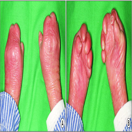
Fig. 1 Preoperative photograph of both hands. Syndactyly of all web space, skin contracture and ulnar deviation of joints are shown. (A) Top view. (B) Bottom view.
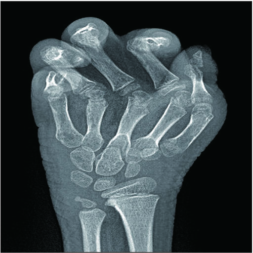
Fig. 2 Preoperative X-ray of right hand. Angle of ulnar deviation of metacarpophalangeal joint of second finger is 60 degree.
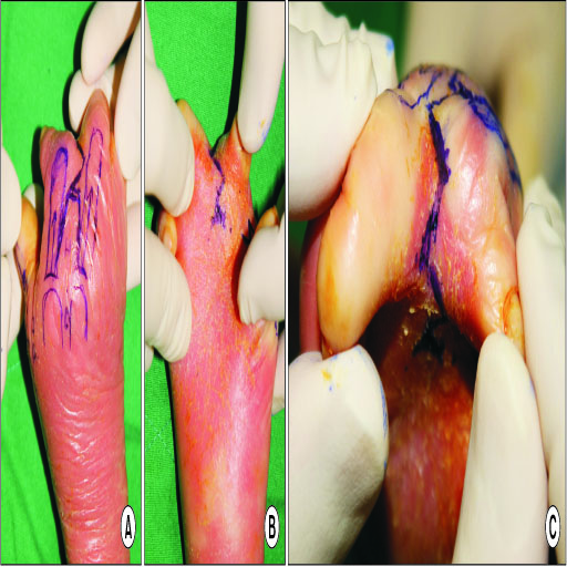
Fig. 3 Photograph of intraoperative design. Zigzag incision, dorsal quadriangular flap and volar triangular flap are drawn. (A) Top view. (B) Bottom view. (C) Tip view.

Fig. 4 Immediate postoperative photograph. Right second web space is fully released with full-thickness skin graft. (A) Top view. (B) Bottom view. (C) Tip view.
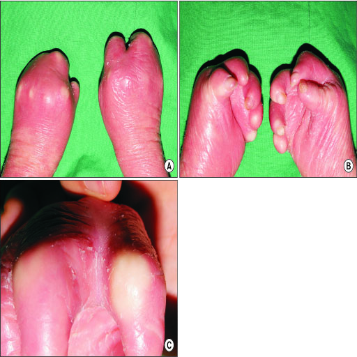
Fig. 5 Postoperative 6-month photograph. Right second web space is well-healed without complication. (A) Top view. (B) Bottom view. (C) Tip view.
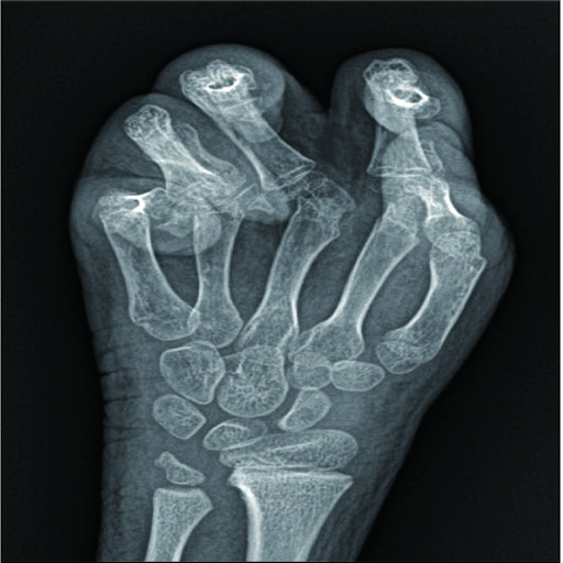
Fig. 6 Postoperative X-ray of right hand. Angle of ulnar deviation of metacarpophalangeal joint of second finger is 25 degree.

Fig. 7 Postoperative wound dehiscence in donor site. At postoperative 2 weeks, left inguinal area underwent minor wound dehiscence which required surgical revision.
Reference
-
1. Kelsell DP, Norgett EE, Unsworth H, et al. Mutations in ABCA12 underlie the severe congenital skin disease harlequin ichthyosis. Am J Hum Genet. 2005; 76:794–803.2. Akiyama M, Sugiyama-Nakagiri Y, Sakai K, et al. Mutations in lipid transporter ABCA12 in harlequin ichthyosis and functional recovery by corrective gene transfer. J Clin Invest. 2005; 115:1777–1784.
Article3. Thomas AC, Tattersall D, Norgett EE, O'Toole EA, Kelsell DP. Premature terminal differentiation and a reduction in specific proteases associated with loss of ABCA12 in Harlequin ichthyosis. Am J Pathol. 2009; 174:970–978.
Article4. Dale BA, Kam E. Harlequin ichthyosis. Variability in expression and hypothesis for disease mechanism. Arch Dermatol. 1993; 129:1471–1477.
Article5. Rajpopat S, Moss C, Mellerio J, et al. Harlequin ichthyosis: a review of clinical and molecular findings in 45 cases. Arch Dermatol. 2011; 147:681–686.6. Toledo LC, Ger E. Evaluation of the operative treatment of syndactyly. J Hand Surg Am. 1979; 4:556–564.
Article7. Vekris MD, Lykissas MG, Soucacos PN, Korompilias AV, Beris AE. Congenital syndactyly: outcome of surgical treatment in 131 webs. Tech Hand Up Extrem Surg. 2010; 14:2–7.
Article8. Werner S, Krieg T, Smola H. Keratinocyte-fibroblast interactions in wound healing. J Invest Dermatol. 2007; 127:998–1008.
Article9. Overland J, Johnstone B. Surgical management of the hand manifestations of harlequin ichthyosis. ANZ J Surg. 2018; DOI: 10.1111/ans.14435. [Epub ahead of print].
Article
