Radiographic Progression of Osteoarthritis after Rotational Acetabular Osteotomy: Minimum 10-Year Follow-up Outcome According to the Tönnis Grade
- Affiliations
-
- 1Department of Orthopaedic Surgery, Keimyung University Dongsan Medical Center, Daegu, Korea. oslee@dsmc.or.kr
- 2Department of Orthopaedic Surgery, Hanmi Hospital, Daegu, Korea.
- KMID: 2418749
- DOI: http://doi.org/10.4055/cios.2018.10.3.299
Abstract
- BACKGROUND
Although satisfactory mid- to long-term results of rotational acetabular osteotomy for early osteoarthritis secondary to acetabular dysplasia have been reported, there is still controversy about the long-term effects of this surgery in more advanced osteoarthritis. The purpose of this study was to investigate the radiographic progression of osteoarthritic changes after rotational acetabular osteotomy in acetabular dysplasia according to the preoperative Tönnis grade and evaluate its effects after minimum 10-year follow-up.
METHODS
We performed 71 consecutive rotational acetabular osteotomies in 64 patients with symptomatic acetabular dysplasia between November 1984 and April 2005. Of these, 46 hips (four hips with Tönnis grade 0, 30 with grade 1, and 12 with grade 2) whose clinical and radiographic findings were available after minimum 10-year follow-up were evaluated in this study. The mean age at the time of surgery was 39.0 years (range, 18 to 62 years) and the average follow-up duration was 17.3 years (range, 10.0 to 27.7 years). Clinical and radiographic evaluations were performed according to the preoperative Tönnis grade.
RESULTS
The average Harris hip score improved from 71.8 (range, 58 to 89) to 85.1 (range, 62 to 98). The radiographic parameters also improved in all Tönnis grades after the index surgery. Although the improvement of radiographic parameters was not different between preoperative Tönnis grades, the incidence of osteoarthritic progression was significantly different between grades (zero in Tönnis grade 0, four in Tönnis grade 1, and 10 in Tönnis grade 2; p < 0.001). The mean age at the time of surgery was also significantly older in osteoarthritic progression patients (p < 0.002). Kaplan-Meier survivorship analysis, with radiographic progression of osteoarthritis as the endpoint, predicted a 10-year survival rate of 100% in Tönnis grade 0, 85.7% in Tönnis grade 1, and 14.3% in Tönnis grade 2 (p < 0.001).
CONCLUSIONS
The outcome of rotational acetabular osteotomy in most hips with Tönnis grade 0 and 1 was satisfactory after an average of 17 years of follow-up. The incidence of osteoarthritic progression was higher in Tönnis grade 2 and older age. Our results support that early joint preserving procedure is essential in the case of symptomatic dysplastic hips.
MeSH Terms
Figure
-
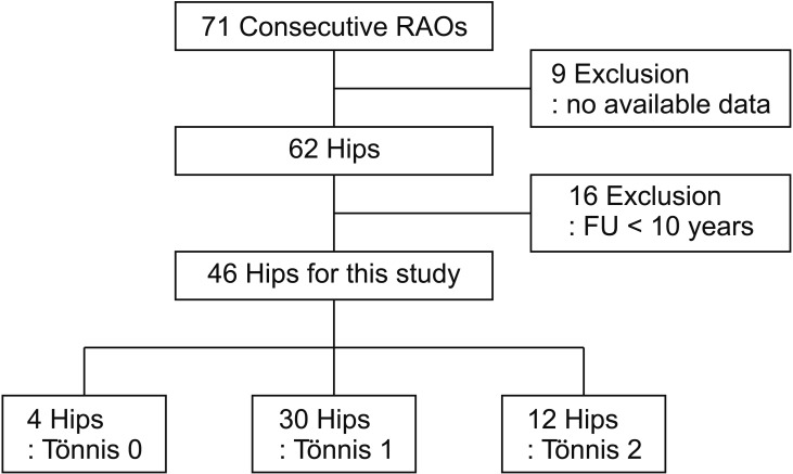
Fig. 1 The flow diagram shows how the final study cohort was determined based on inclusion and exclusion criteria. RAO: rotational acetabular osteotomy, FU: follow-up.
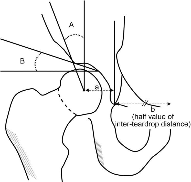
Fig. 2 Radiographic parameters for evaluation of the hip. A: Center-edge angle. B: Acetabular roof angle. The head lateralization index is calculated using the distance between the teardrop and the center of the femoral head (a) divided by the half value of the inter-teardrop distance (b).
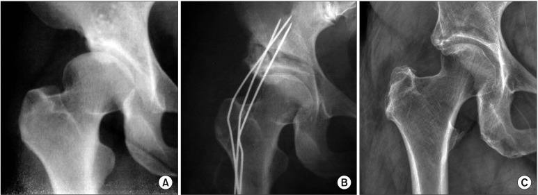
Fig. 3 Anteroposterior radiographs of the right hip joint in a 47-year-old male. (A) The preoperative radiograph shows mild acetabular dysplasia with no sign of degenerative change (Tönnis grade 0). (B) The postoperative radiograph shows improvement of acetabular coverage over the femoral head. Note that the osteotomized fragment and grafted bone were fixed with three Kirschner wires. (C) The Radiograph obtained 26 years after rotational acetabular osteotomy shows maintenance of good coverage and no progression of osteoarthritis.
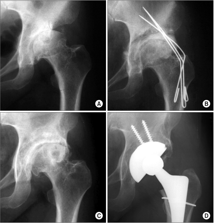
Fig. 4 Anteroposterior radiographs of the left hip joint in a 47-year-old female. (A) The preoperative radiograph shows moderate joint space narrowing with subchondral cyst in the femoral head (Tönnis grade 2). (B) The postoperative radiograph shows improvement of acetabular coverage over the femoral head. (C) The postoperative radiograph obtained 7 years after rotational acetabular osteotomy shows progression of osteoarthritis. (D) Total hip arthroplasty was performed 7.5 years after the index surgery due to progressive osteoarthritis.
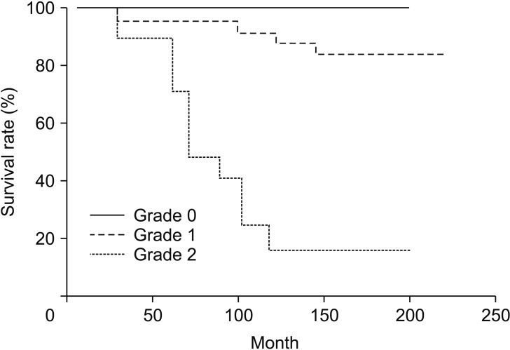
Fig. 5 The Kaplan-Meier survivorship analysis demonstrates the cumulative probability of osteoarthritic progression according to the Tönnis grade.
Reference
-
1. Dorey FJ. Survivorship analysis of surgical treatment of the hip in young patients. Clin Orthop Relat Res. 2004; (418):23–28.
Article2. Yasunaga Y, Fujii J, Tanaka R, et al. Rotational acetabular osteotomy. Clin Orthop Surg. 2017; 9(2):129–135. PMID: 28567213.
Article3. Hasegawa Y, Iwata H, Mizuno M, Genda E, Sato S, Miura T. The natural course of osteoarthritis of the hip due to subluxation or acetabular dysplasia. Arch Orthop Trauma Surg. 1992; 111(4):187–191. PMID: 1622705.
Article4. Hartofilakidis G, Karachalios T, Zacharakis N. Charnley low friction arthroplasty in young patients with osteoarthritis: a 12- to 24-year clinical and radiographic followup study of 84 cases. Clin Orthop Relat Res. 1997; (341):51–54. PMID: 9269154.
Article5. Steel HH. Triple osteotomy of the innominate bone. J Bone Joint Surg Am. 1973; 55(2):343–350. PMID: 4572223.
Article6. Eppright RH. Dial osteotomy of the acetabulum in the treatment of dysplasia of the hip. J Bone Joint Surg Am. 1975; 57(8):1172.7. Wagner H. Osteotomies for congenital hip dislocation. The Hip Society. The hip: Proceedings of the 4th Open Scientific Meeting of the Hip Society. St Louis, MO: C.V. Mosby;1976. p. 45–66.8. Ninomiya S, Tagawa H. Rotational acetabular osteotomy for the dysplastic hip. J Bone Joint Surg Am. 1984; 66(3):430–436. PMID: 6699061.
Article9. Ganz R, Klaue K, Vinh TS, Mast JW. A new periacetabular osteotomy for the treatment of hip dysplasias: technique and preliminary results. Clin Orthop Relat Res. 1988; (232):26–36.
Article10. Lee CB, Millis MB. Patient selection for rotational pelvic osteotomy. Instr Course Lect. 2013; 62:265–277. PMID: 23395032.11. de Kleuver M, Kooijman MA, Pavlov PW, Veth RP. Triple osteotomy of the pelvis for acetabular dysplasia: results at 8 to 15 years. J Bone Joint Surg Br. 1997; 79(2):225–229. PMID: 9119847.12. Nakamura S, Ninomiya S, Takatori Y, Morimoto S, Umeyama T. Long-term outcome of rotational acetabular osteotomy: 145 hips followed for 10–23 years. Acta Orthop Scand. 1998; 69(3):259–265. PMID: 9703399.
Article13. Schramm M, Pitto RP, Rohm E, Hohmann D. Long-term results of spherical acetabular osteotomy. J Bone Joint Surg Br. 1999; 81(1):60–66. PMID: 10068005.
Article14. Nozawa M, Shitoto K, Matsuda K, Maezawa K, Kurosawa H. Rotational acetabular osteotomy for acetabular dysplasia: a follow-up for more than ten years. J Bone Joint Surg Br. 2002; 84(1):59–65. PMID: 11837834.15. Nozawa M, Maezawa K, Matsuda K, Kim S, Shitoto K, Kurosawa H. Rotational acetabular osteotomy for advanced osteoarthritis of the hip joint with acetabular dysplasia. Int Orthop. 2009; 33(6):1549–1553. PMID: 18853158.
Article16. Kaneuji A, Sugimori T, Ichiseki T, Fukui K, Takahashi E, Matsumoto T. Rotational acetabular osteotomy for osteoarthritis with acetabular dysplasia: conversion rate to total hip arthroplasty within twenty years and osteoarthritis progression after a minimum of twenty years. J Bone Joint Surg Am. 2015; 97(9):726–732. PMID: 25948519.17. Tonnis D. Congenital dysplasia and dislocation of the hip in children and adults. New York, NY: Springer-Verlag Berlin Heidelberg;1987. p. 165–171.18. Wiberg G. Studies on dysplastic acetabula and congenital subluxation of the hip joint: with special reference to the complication of osteoarthritis. Acta Chir Scand. 1939; 83(Suppl 58):53–68.19. Massie WK, Howorth MB. Congenital dislocation of the hip. Part I: method of grading results. J Bone Joint Surg Am. 1950; 32(3):519–531. PMID: 15428474.20. Ninomiya S. Rotational acetabular osteotomy for the severely dysplastic hip in the adolescent and adult. Clin Orthop Relat Res. 1989; (247):127–137. PMID: 2791382.
Article21. Murphy SB, Kijewski PK, Millis MB, Harless A. Acetabular dysplasia in the adolescent and young adult. Clin Orthop Relat Res. 1990; (261):214–223. PMID: 2245547.
Article22. Takatori Y, Ninomiya S, Nakamura S, et al. Long-term results of rotational acetabular osteotomy in patients with slight narrowing of the joint space on preoperative radiographic findings. J Orthop Sci. 2001; 6(2):137–140. PMID: 11484099.
Article23. Yasunaga Y, Ochi M, Yamasaki T, Shoji T, Izumi S. Rotational acetabular osteotomy for pre- and early osteoarthritis secondary to dysplasia provides durable results at 20 years. Clin Orthop Relat Res. 2016; 474(10):2145–2153. PMID: 27121873.
Article24. Okano K, Enomoto H, Osaki M, Shindo H. Outcome of rotational acetabular osteotomy for early hip osteoarthritis secondary to dysplasia related to femoral head shape: 49 hips followed for 10–17 years. Acta Orthop. 2008; 79(1):12–17. PMID: 18283566.
Article25. Yasunaga Y, Ochi M, Terayama H, Tanaka R, Yamasaki T, Ishii Y. Rotational acetabular osteotomy for advanced osteoarthritis secondary to dysplasia of the hip. J Bone Joint Surg Am. 2006; 88(9):1915–1919. PMID: 16951105.
Article26. Yamaguchi J, Hasegawa Y, Kanoh T, Seki T, Kawabe K. Similar survival of eccentric rotational acetabular osteotomy in patients younger and older than 50 years. Clin Orthop Relat Res. 2009; 467(10):2630–2637. PMID: 19424675.
Article27. Yasunaga Y, Takahashi K, Ochi M, et al. Rotational acetabular osteotomy in patients forty-six years of age or older: comparison with younger patients. J Bone Joint Surg Am. 2003; 85(2):266–272. PMID: 12571304.
- Full Text Links
-
- Actions
-
Cited
- CITED
-
- Close
- Share
-
- Similar articles
-
- Rotational Acetabular Osteotomy for the Dysplastic Hip: A Follow-up for 5 to 18 years
- Pelvic Osteotomy in Adults
- Periacetabular Rotational Osteotomy in Hip Dysplasia: Short Term Follow up Result
- Rotational Acetabular Osteotomy for the Dysplastic Acetabulum
- Arthroscopic Repair of Acetabular Labral Tears Associated with Femoroacetabular Impingement: 7–10 Years of Long-Term Follow-up Results
