Korean J Gastroenterol.
2018 Jul;72(1):33-36. 10.4166/kjg.2018.72.1.33.
Anisakiasis Induced Segmental Jejunum Obstruction
- Affiliations
-
- 1Department of Internal Medicine, Maryknoll Hospital, Busan, Korea. bumhee7@hanmail.net
- 2Department of General surgery, Maryknoll Hospital, Busan, Korea.
- 3Department of Radiology, Maryknoll Hospital, Busan, Korea.
- 4Department of Pathology, Maryknoll Hospital, Busan, Korea.
- KMID: 2416934
- DOI: http://doi.org/10.4166/kjg.2018.72.1.33
Abstract
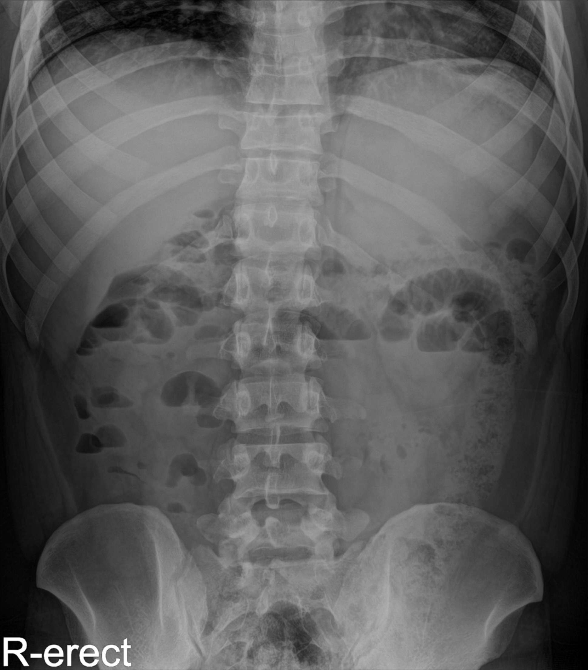
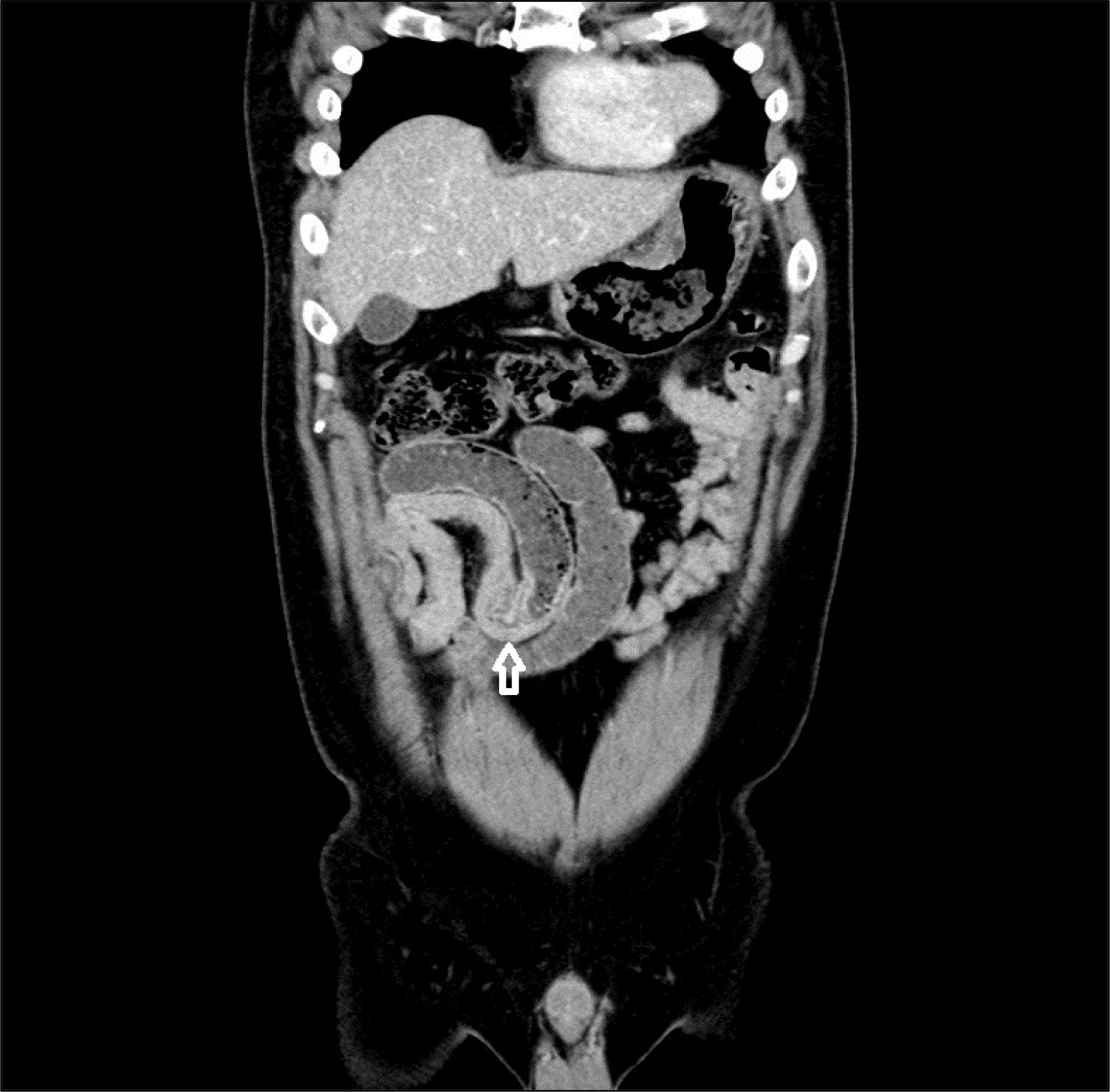
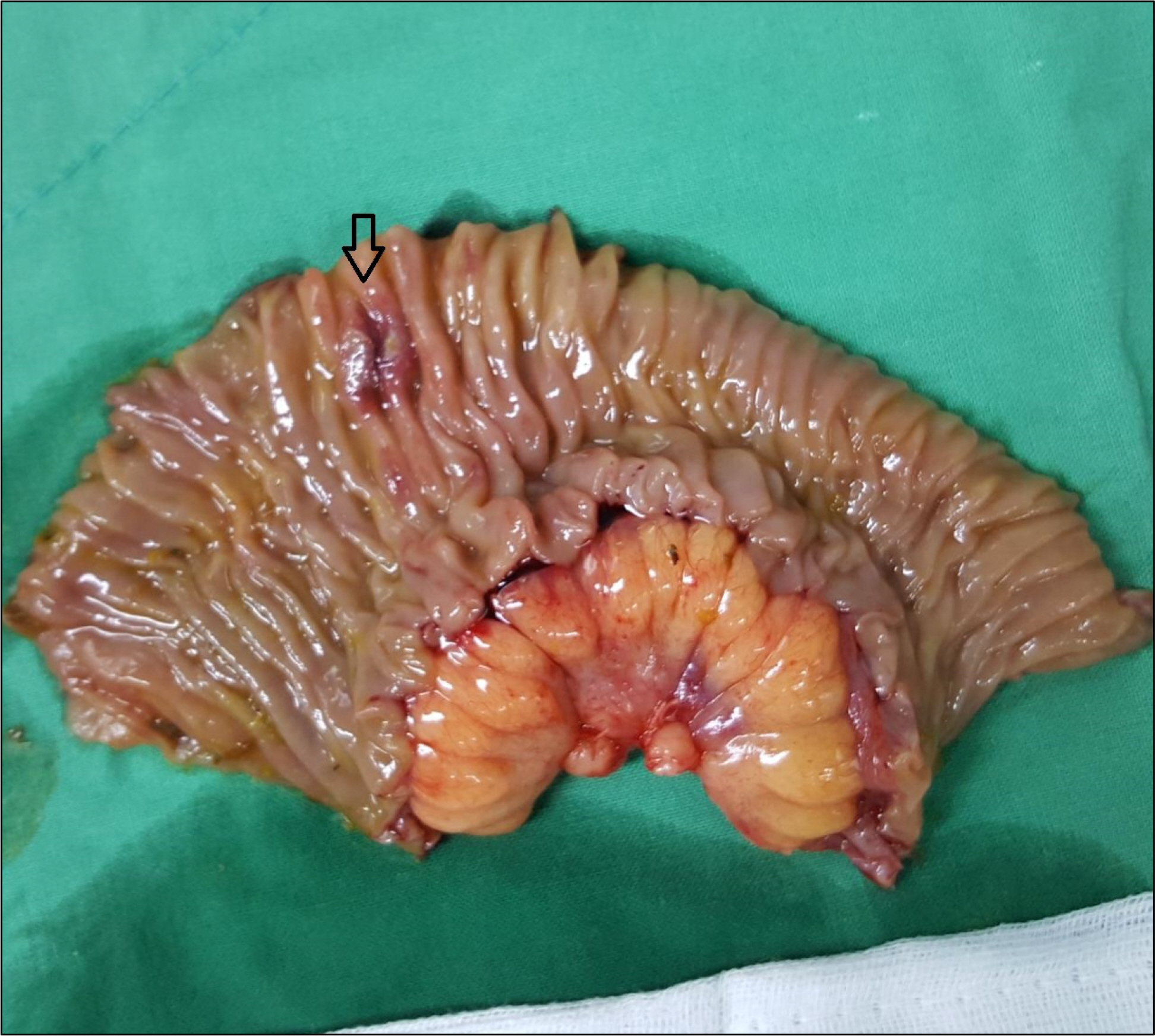
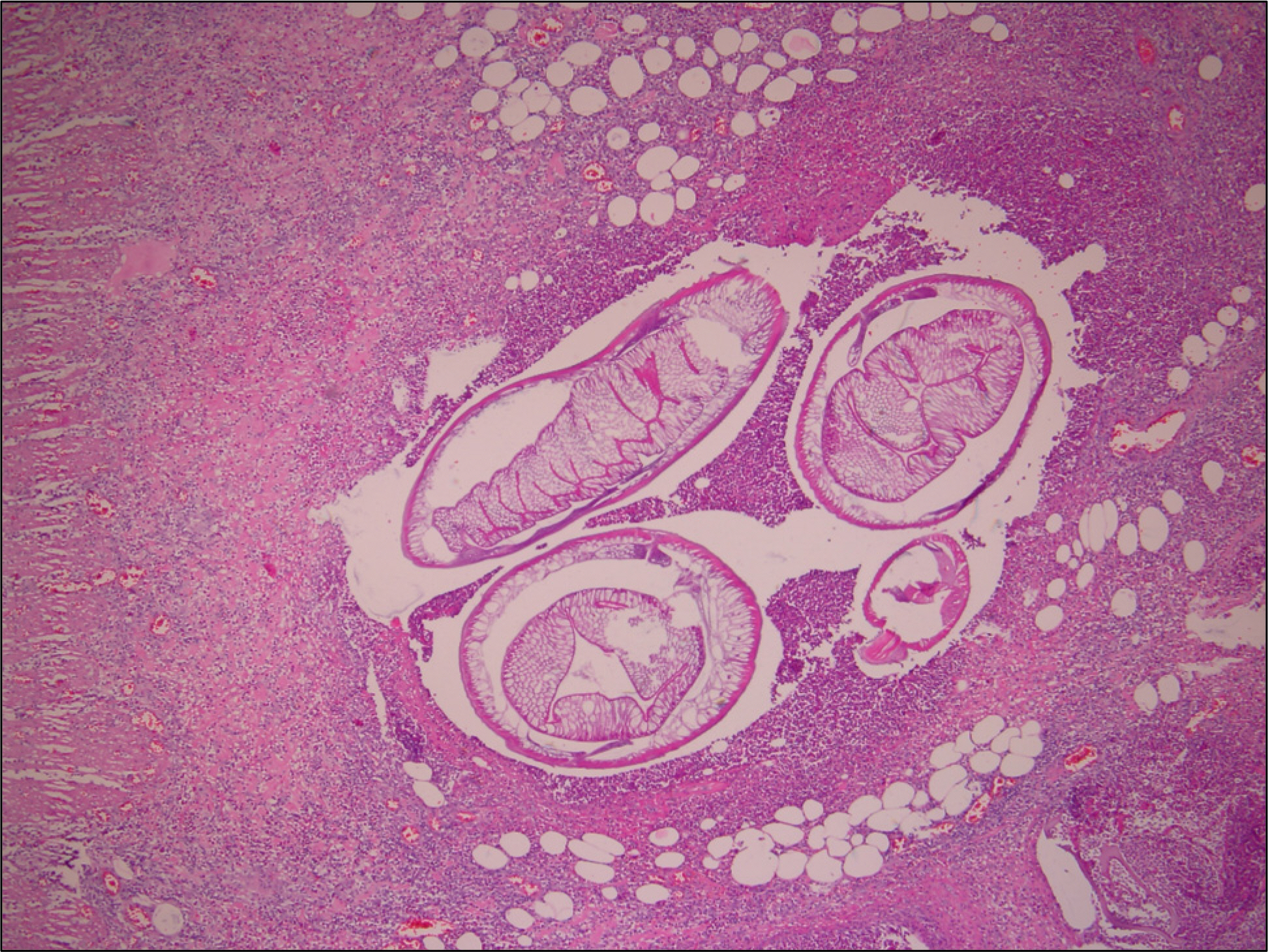
- Human anisakiasis is a disease caused by an infestation of the third stage larvae of family anisakidae. The ingested larvae invade the gastrointestinal wall, causing clinical symptoms that include abdomen pain, nausea, and vomiting. Although enteric anisakiasis is extremely rare, it can induce intestinal obstruction. We report a case in which emergency surgery was needed due to intestinal obstruction that coincided with symptoms related to anisakiasis, along with a brief literature review.
Keyword
Figure
Reference
-
References
1. Seo BS. Clinical Parasitology. 3rd ed.Seoul: Ilchokak;1997.2. van Thiel P, Kuipers FC, Roskam RT. A nematode parasitic to herring, causing acute abdominal syndromes in man. Trop Geogr Med. 1960; 12:97–113.3. Van Thiel PH. The present state of anisakiasis and its causative worms. Trop Geogr Med. 1976; 28:75–85.4. Kim CH, Chung BS, Moon YI, Chun SH. A case report on human infection with Anisakis sp. in Korea. Korean J Parasitol. 1971; 9:39–43.
Article5. Lee KH, Koo JT, Song JH, Hyun MS, Jhi CJ. Acute gastric anisa-kiasis-endoscopic, radiologic diagnosis and its management. Korean J Med. 1981; 24:1220–1227.6. Han DS, Han YB, Park DL, Kim SH, Kim SS. Clinical study of anisakiasis. J Korean Med Assoc. 1988; 31:645.7. Chapman HB, Daniel HC. Pathology of tropical and extraordinary disease. Volume 2. 2nd ed.Washington, D.C: Armed Forces Institute of Pathology;1976.8. Park SH, Suh JM, Shim KS, Baeg NJ, Kim BS, Moon IS. A case report of intestinal anisakiasis. Korean J Gastrointest Endosc. 1990; 10:373–375.9. Pinkus GS, Collidge C, Little MD. Intestinal anisakiasis. first case report from North America. Am J Med. 1975; 59:114–120.10. Ishikura H, Kikuchi K. Intestinal anisakiasis in japan: infected fish, sero-immunological diagnosis, and prevention. 2nd ed.Tokyo: Springer-Verlag Berlin and Heidelberg;1990.11. Takano Y, Gomi K, Endo T, et al. Small intestinal obstruction caused by anisakiasis. Case Rep Infect Dis. 2013; 2013:401937.
Article12. Kojima G, Usuki S, Mizokami K, Tanabe M, Machi J. Intestinal anisakiasis as a rare cause of small bowel obstruction. Am J Emerg Med. 2013; 31:1422. e1-e2.
Article13. Shrestha S, Kisino A, Watanabe M, et al. Intestinal anisakiasis treated successfully with conservative therapy: importance of clinical diagnosis. World J Gastroenterol. 2014; 20:598–602.
Article14. Matsuo S, Azuma T, Susumu S, Yamaguchi S, Obata S, Hayashi T. Small bowel anisakiasis: a report of two cases. World J Gastroenterol. 2006; 12:4106–4108.
- Full Text Links
- Actions
-
Cited
- CITED
-
- Close
- Share
- Similar articles
-
- Small Bowel Obstruction Caused by Acute Invasive Enteric Anisakiasis
- A case of anisakiasis causing intestinal obstruction
- Intestinal Obstruction Caused by Anisakiasis
- Duodenal Anisakiasis Presenting as Bowel Obstruction and Fistula Formation: A Case Report
- A Case of Small Bowel Obstruction and Perforation by Anisakiasis


