Central Nervous System Involvement in a Patient with Multiple Myeloma Manifesting as an Intraventricular Mass with Leptomeningeal Spread
- Affiliations
-
- 1Department of Radiology, Dongguk University Ilsan Hospital, Goyang, Korea.
- 2Department of Laboratory Medicine, Dongguk University Ilsan Hospital, Goyang, Korea.
- 3Division of Hematology and Medical Oncology, Department of Internal Medicine, Dongguk University Ilsan Hospital, Goyang, Korea. altvega@gmail.com
- KMID: 2416406
- DOI: http://doi.org/10.3348/jksr.2018.79.1.50
Abstract
- Central nervous system involvement in multiple myeloma (CNS-MM) is a rare condition. Various manifestations of CNS-MM have been reported, including dural, parenchymal, and leptomeningeal involvement. Among them, leptomeningeal involvement is less common and intraventricular involvement is exceptional, with only one case reported in the literature. Herein, we report the first case of CNS-MM manifesting as an intraventricular mass with leptomeningeal involvement combined with perineural spread. We also describe characteristic computed tomography and magnetic resonance imaging findings of intraventricular multiple myeloma.
MeSH Terms
Figure
-
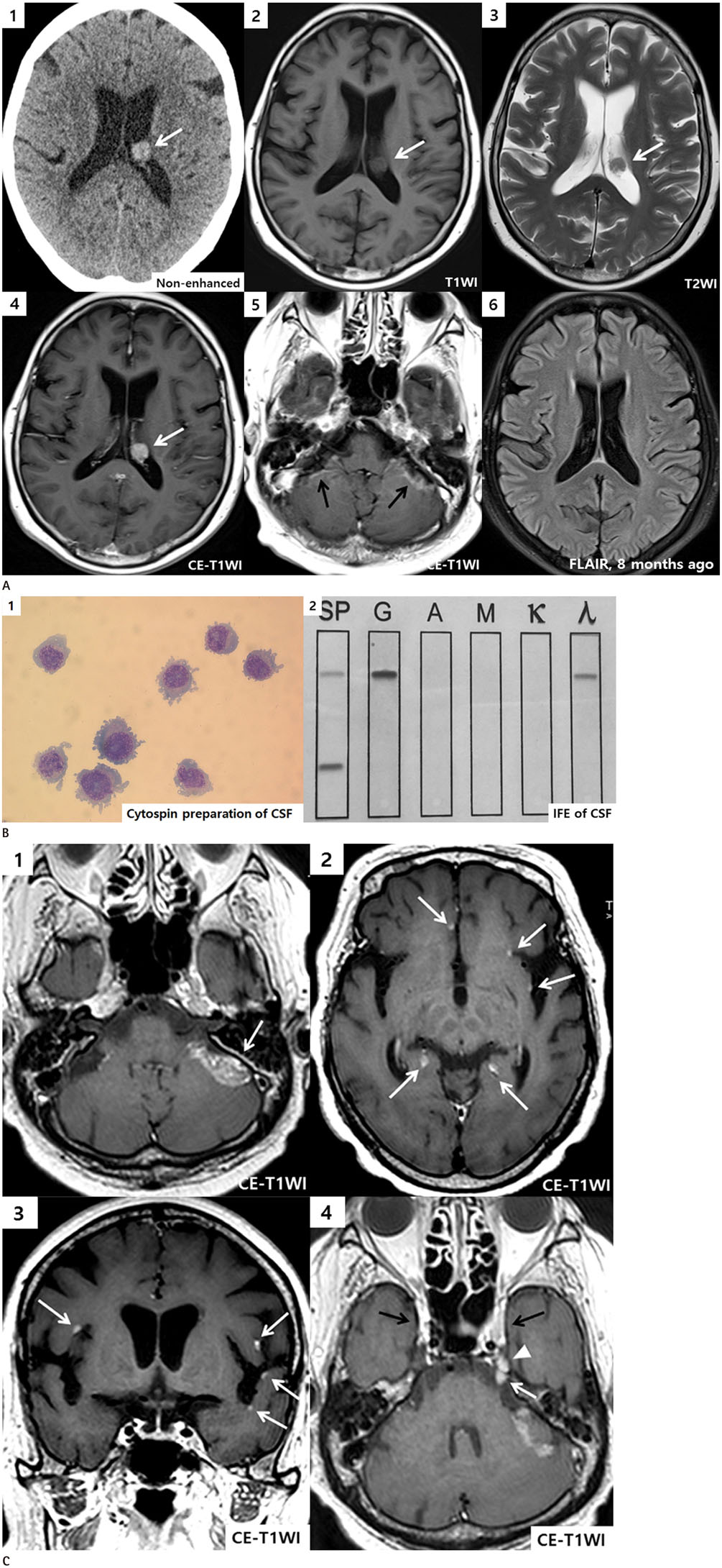
Fig. 1 A 66-year-old female with central nervous system involvement of multiple myeloma manifesting as an intraventricular mass with leptomeningeal spread. A. Non-CE CT scan shows a small ovoid hyperdense mass (50 Hounsfield units) (arrow) in trigone of left lateral ventricle (1). Iso-signal intense mass (arrows) is detected on both T1WI and T2WI (2, 3). CE T1WIs present strong homogeneous enhancement of intraventricular mass (white arrow) and multifocal intermittent leptomeningeal enhancement (black arrows), especially along the left cerebellar folia (4, 5). This intraventricular mass is not revealed in previous FLAIR image performed 8 months ago (6). CE = contrast-enhanced, FLAIR = fluid-attenuated inversion recovery, T1WI = T1-weighted image, T2WI = T2-weighted image B. Cytospin preparation of CSF from a MM patient with leptomeningeal involvement shows abundant malignant plasma cells (Wright stain, × 1000) (1), IFE of CSF demonstrates a monoclonal gammopathy, IgG and lambda type (2). CSF = cerebrospinal fluid, IFE = immunofixation electrophoresis, MM = multiple myeloma C. Follow up MRI of CE T1WI (2 weeks later) demonstrates increased size of leptomeningeal enhancing lesion (arrow) in the posterior fossa (1), multifocal leptomeningeal enhancing nodules (arrows) are noted (2, 3), abnormal enhancement along the cisternal segment of left trigeminal nerve (white arrow) is also noted with focal extension into ipsilateral Meckel's cave (arrowhead) and both cavernous sinuses show bulging contour with strong enhancement (black arrows) (4). These image findings suggest leptomeningeal involvement of MM with perineural spread. CE = contrast-enhanced, MM = multiple myeloma, T1WI = T1-weighted image
Reference
-
1. Palumbo A, Anderson K. Multiple myeloma. N Engl J Med. 2011; 364:1046–1060.
Article2. Lasocki A, Gangatharan S, Gaillard F, Harrison SJ. Intracranial involvement by multiple myeloma. Clin Radiol. 2015; 70:890–897.
Article3. Gozzetti A, Cerase A, Lotti F, Rossi D, Palumbo A, Petrucci MT, et al. Extramedullary intracranial localization of multiple myeloma and treatment with novel agents: a retrospective survey of 50 patients. Cancer. 2012; 118:1574–1584.
Article4. Méndez CE, Hwang BJ, Destian S, Mazumder A, Jagannath S, Vesole DH. Intracranial multifocal dural involvement in multiple myeloma: case report and review of the literature. Clin Lymphoma Myeloma Leuk. 2010; 10:220–223.
Article5. Cerase A, Tarantino A, Gozzetti A, Muccio CF, Gennari P, Monti L, et al. Intracranial involvement in plasmacytomas and multiple myeloma: a pictorial essay. Neuroradiology. 2008; 50:665–674.
Article6. Eum JH, Jeibmann A, Wiesmann W, Paulus W, Ebel H. Multiple myeloma manifesting as an intraventricular brain tumor. J Neurosurg. 2009; 110:737–739.
Article7. Abdallah AO, Atrash S, Shahid Z, Jameel M, Grazziutti M, Apewokin S, et al. Patterns of central nervous system involvement in relapsed and refractory multiple myeloma. Clin Lymphoma Myeloma Leuk. 2014; 14:211–214.
Article8. Seo JM, Lee KS, Yi CA, Kim SH, Park BK, Han BK, et al. A pictorial review on extraosseous manifestations of multiple myelomas. J Korean Soc Radiol. 2011; 64:567–575.
Article9. de la Fuente J, Prieto I, Albo C, Sopeña B, Somolinos N, Martínez C. Plasma cell myeloma presented as myelomatous meningitis. Eur J Haematol. 1994; 53:244–245.10. Patriarca F, Zaja F, Silvestri F, Sperotto A, Scalise A, Gigli G, et al. Meningeal and cerebral involvement in multiple myeloma patients. Ann Hematol. 2001; 80:758–762.
Article
- Full Text Links
-
- Actions
-
Cited
- CITED
-
- Close
- Share
-
- Similar articles
-
- A Case of Multiple Myeloma with Brain Parenchyme Involvement
- Cysticercosis of the Central Nervous System
- Meningeal and Cerebral Involvement of a Plasmacytoma in an IgG Multiple Myeloma Patient: Case Report
- Liver Involvement of Multiple Myeloma Mimicking Intrahepatic Cholangiocarcinoma: A Case Report
- A Rare Case of Diffuse Pachymeningeal Involvement of Multiple Myeloma
