A Case of Multiple Myeloma with Brain Parenchyme Involvement
- Affiliations
-
- 1Department of Neurosurgery, Dongtan Sacred Heart Hospital, College of Medicine, Hallym University, Hwaseong, Korea. troias@naver.com
- 2Department of Pathology, Dongtan Sacred Heart Hospital, College of Medicine, Hallym University, Hwaseong, Korea.
- KMID: 2048484
- DOI: http://doi.org/10.14791/btrt.2013.1.2.103
Abstract
- Multiple myelomas (MM) are characterized by monoclonal proliferation of immunoglobulin (Ig)-secreting plasma cells. Central nervous system involvement is a rare complication of MM, and it can present as either an intraparenchymal or a leptomeningeal lesion. A 77-year-old woman was transferred from the dementia clinic in July 2012. She had a large heterogeneous signal mass with central necrosis and with pial involvement in the left frontal lobe with destruction of the frontal bone that was observed on computed tomography and magnetic resonance imaging. Multiple punched out radiolucent lesions were also noted on the skull X-ray. Serum protein electrophoresis revealed an IgA-kappa monoclonal gammopathy. External lumbar drainage was helpful for treating the fluid collection underneath the scalp after an orbitozygomatic craniotomy with duroplasty.
Keyword
MeSH Terms
Figure
-
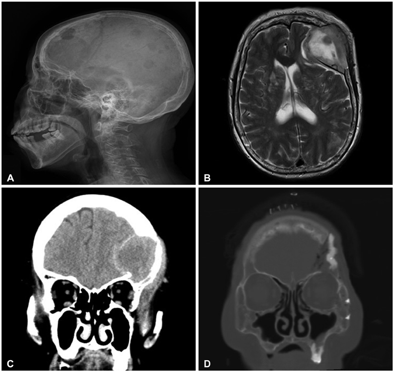
Fig. 1 Image study of the patient. A: Skull X-ray. Multiple punch out lesion was noted. B: T2 weighted magnetic resonance image (axial). Large heterogeneous signal mass with central necrosis and pial involvement is noted in left frontal lobe. Adjacent bony destruction and parenchymal edema are also noted. After contrast media injection, the mass shows strong enhancement. And multiple other smaller well-enhancing nodular or lobulating contoured nodules in both frontal and parietal bones are noted. C: Pre-operation brain computed tomography (CT). Relatively wall marginated bone destruction is noted in left frontal and parietal bones. D: Post-operation brain CT (bone setting image). Post-operative state, left frontal tumor removal and cranioplasty state with orbitozygomatic approach.

Fig. 2 Electrophoresis and Immunofixations. A: Serum electrophoresis. B: Urine electrophoresis. C: Serum Immunofixation. D: Urine Immunofixation, Serum and urine protein electrophoresis revealed monoclonal gammopathy, IgA kappa type, free kappa light chains.
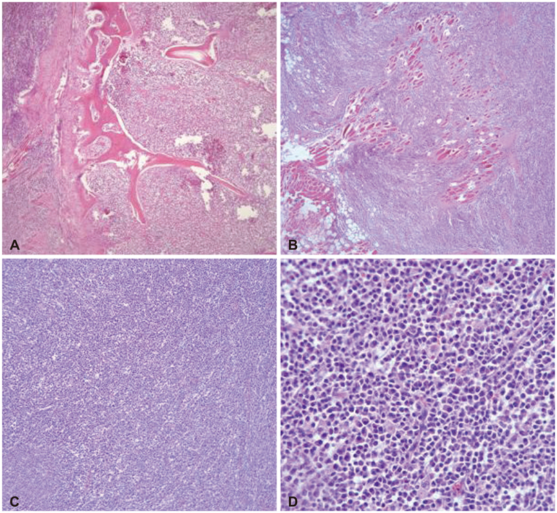
Fig. 3 The biopsy shows diffuse infiltration of atypical plasma cell with prominent nucleoli and mitosis at bone, skeletal muscle, brain parenchyma. A: Bone involvement. B: Skeletal muscle involvement. C: Neoplastic cell (H&E, ×40). D: Neoplastic cell (H&E,×400).
Reference
-
1. Kyle RA. Multiple myeloma: review of 869 cases. Mayo Clin Proc. 1975; 50:29–40.2. Turhal N, Henehan MD, Kaplan KL. Multiple myeloma: a patient with unusual features including intracranial and meningeal involvement, testicular involvement, organomegaly, and plasma cell leukemia. Am J Hematol. 1998; 57:51–56.
Article3. Somers LJ, Shaw B, Lyn BE, McMillan AM, Mahendra P. Meningeal myeloma in the absence of systemic disease, and as the initial feature of disease progression. Clin Lab Haematol. 1998; 20:189–190.
Article4. Peest D. [Multiple myeloma]. Ther Umsch. 1996; 53:147–151.5. Fassas AB, Muwalla F, Berryman T, et al. Myeloma of the central nervous system: association with high-risk chromosomal abnormalities, plasmablastic morphology and extramedullary manifestations. Br J Haematol. 2002; 117:103–108.
Article6. Roddie P, Collie D, Johnson P. Myelomatous involvement of the dura mater: a rare complication of multiple myeloma. J Clin Pathol. 2000; 53:398–399.
Article7. Méndez CE, Hwang BJ, Destian S, Mazumder A, Jagannath S, Vesole DH. Intracranial multifocal dural involvement in multiple myeloma: case report and review of the literature. Clin Lymphoma Myeloma Leuk. 2010; 10:220–223.
Article8. Paul RH, Piatt AL, Whelihan WM, Malloy PF. Neuropsychological and magnetic resonance imaging abnormalities associated with a plasmacytoma of the frontal dura: a case report. Neuropsychiatry Neuropsychol Behav Neurol. 2000; 13:143–147.9. Cerase A, Tarantino A, Gozzetti A, et al. Intracranial involvement in plasmacytomas and multiple myeloma: a pictorial essay. Neuroradiology. 2008; 50:665–674.
Article10. Petersen SL, Wagner A, Gimsing P. Cerebral and meningeal multiple myeloma after autologous stem cell transplantation. A case report and review of the literature. Am J Hematol. 1999; 62:228–233.
Article
- Full Text Links
-
- Actions
-
Cited
- CITED
-
- Close
- Share
-
- Similar articles
-
- Meningeal and Cerebral Involvement of a Plasmacytoma in an IgG Multiple Myeloma Patient: Case Report
- Liver Involvement of Multiple Myeloma Mimicking Intrahepatic Cholangiocarcinoma: A Case Report
- A Case of Intracranial Involvement in Plasma Cell Myeloma
- A Rare Case of Diffuse Pachymeningeal Involvement of Multiple Myeloma
- A Case of Maxillary Sinus Involvement in Multiple Myeloma
