J Korean Assoc Oral Maxillofac Surg.
2018 Jun;44(3):136-139. 10.5125/jkaoms.2018.44.3.136.
Angioleiomyoma of the oral cavity: a case report and brief review of the literature
- Affiliations
-
- 1Department of Oral Medicine and Pathology, Periodontology and Implant Biology, School of Dentistry, Aristotle University of Thessaloniki, Thessaloniki, Greece. amatiakis@dent.auth.gr
- 2Department of Preventive Dentistry, Periodontology and Implant Biology, School of Dentistry, Aristotle University of Thessaloniki, Thessaloniki, Greece.
- 3Pathologist, Thessaloniki, Greece.
- KMID: 2414433
- DOI: http://doi.org/10.5125/jkaoms.2018.44.3.136
Abstract
- This study presents a case of an oral angioleiomyoma along with its clinical diagnostic approach and laboratory confirmation. The differential diagnosis, especially from angioleiomyosarcoma, is also included. A 51-year-old patient presented with a tumor-like lesion on his upper labial mucosa. The clinical examination revealed a benign lesion that was surgically removed. Histopathological and immunohistochemical examinations confirmed the diagnosis of an oral angioleiomyoma. The post-surgical period was uneventful. No recurrence had occurred after a year of follow-up surveillance. Oral angioleiomyoma is a very rarely occurring oral lesion. Clinically, it may mimic some benign lesions, including fibroma, pyogenic granuloma or minor salivary gland tumor. Surgical excision is the treatment of choice. Histological and immunohistochemical examination can confirm the diagnosis. The differential diagnosis is crucial to rule out angioleiomyosarcoma.
MeSH Terms
Figure
-
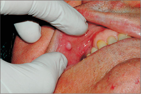
Fig. 1 Initial clinical appearance.
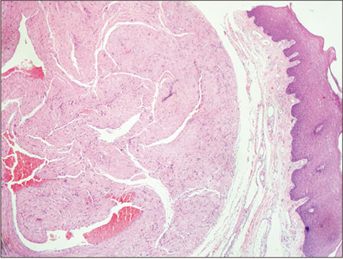
Fig. 2 Bundles of smooth muscle fibers of thick-walled blood vessels (H&E staining, ×40).
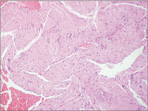
Fig. 3 Similar microscopic findings with no nuclear atypia or mitoses (H&E staining, ×400).
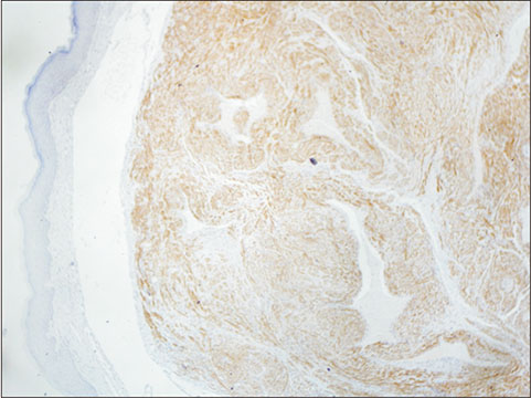
Fig. 4 Positive immunohistochemical reaction of the smooth muscle fibers (h-caldesmon staining, ×40).
Reference
-
1. BBaden E, Doyle JL, Lederman DA. Leiomyoma of the oral cavity: a light microscopic and immunohistochemical study with review of the literature from 1884 to 1992. Eur J Cancer B Oral Oncol. 1994; 30B:1–7.2. Brooks JK, Nikitakis NG, Goodman NJ, Levy BA. Clinicopathologic characterization of oral angioleiomyomas. Oral Surg Oral Med Oral Pathol Oral Radiol Endod. 2002; 94:221–227.
Article3. Liu Y, Li B, Li L, Liu Y, Wang C, Zha L. Angioleiomyomas in the head and neck: a retrospective clinical and immunohistochemical analysis. Oncol Lett. 2014; 8:241–247.
Article4. Hassona Y, Sawair F, Scully C. Angioleiomyoma of the upper lip. BMJ Case Rep. 2017; DOI: 10.1136/bcr-2016-219172.
Article5. Epivatianos A, Trigonidis G, Papanayotou P. Vascular leiomyoma of the oral cavity. J Oral Maxillofac Surg. 1985; 43:377–382.
Article6. Ishikawa S, Fuyama S, Kobayashi T, Taira Y, Sugano A, Iino M. Angioleiomyoma of the tongue: a case report and review of the literature. Odontology. 2016; 104:119–122.
Article7. Osano H, Ioka Y, Okamoto R, Nakai Y, Hayashi H, Tsuchiya Y, et al. Angioleiomyoma of the cheek: a case report. J Oral Sci. 2015; 57:63–66.
Article8. Eley KA, Alroyayamina S, Golding SJ, Tiam RN, Watt-Smith SR. Angioleiomyoma of the hard palate: report of a case and review of the literature and magnetic resonance imaging findings of this rare entity. Oral Surg Oral Med Oral Pathol Oral Radiol. 2012; 114:e45–e49.
Article9. Tsuji T, Satoh K, Nakano H, Kogo M. Clinical characteristics of angioleiomyoma of the hard palate: report of a case and an analysis of the reported cases. J Oral Maxillofac Surg. 2014; 72:920–926.
Article10. Rawal SY, Rawal YB. Angioleiomyoma (vascular leiomyoma) of the oral cavity. Head Neck Pathol. 2018; 12:123–126.
Article11. Ranjan S, Singh KT. Gingival angioleiomyoma-infrequent lesion of oral cavity at a rare site. J Oral Maxillofac Pathol. 2014; 18:107–110.
Article12. Arpağ OF, Damlar I, Kılıç S, Altan A, Taş ZA, Özgür T. Angioleiomyoma of the gingiva: a report of two cases. J Korean Assoc Oral Maxillofac Surg. 2016; 42:115–119.
Article13. Montague LJ, Fitzpatrick SG, Islam NM, Cohen DM, Bhattacharyya I. Extensively ossifying oral leiomyoma: a rare histologic finding. Head Neck Pathol. 2014; 8:311–316.
Article14. Bajpai M, Pardhe N, Kumar M. Angioleiomyoma of gingiva masquerading as pyogenic granuloma. J Coll Physicians Surg Pak. 2016; 26:631–632.15. Nikitakis NG, Lopes MA, Bailey JS, Blanchaert RH Jr, Ord RA, Sauk JJ. Oral leiomyosarcoma: review of the literature and report of two cases with assessment of the prognostic and diagnostic significance of immunohistochemical and molecular markers. Oral Oncol. 2002; 38:201–208.
Article
