Deep anterior lamellar keratoplasty of dog eyes using the big-bubble technique
- Affiliations
-
- 1Department of Veterinary Clinical Science, College of Veterinary Medicine, Seoul National University, Seoul 08826, Korea. kmseo@snu.ac.kr
- 2Royal Animal Medical Center, Seoul 02117, Korea.
- KMID: 2413134
- DOI: http://doi.org/10.4142/jvs.2016.17.3.347
Abstract
- This study was conducted to establish the feasibility of corneal transplantation using the big-bubble technique (BBT) to perform deep anterior lamellar keratoplasty (DALK) in three dogs. After the cornea was trephined 750 µm, 4 mL of air was injected, and the blanched stroma was removed to expose Descemet's membrane (DM). The donor corneal button, which was gently stripped off the DM, was sutured onto the bare DM of the recipient cornea. The dogs received topical antibiotics every 6 h for 7 days and 2% cyclosporine ointment every 12 h for 1 month. The eyes were examined post-operatively at 7, 14, 21, 28 and 150 days. The central portion of the transplanted cornea stayed transparent while corneal haze developed around the transplanted margin. Menace response was normal even though the transplanted cornea was edematous until 3 weeks after surgery. A marginal haze was rarely observed between the donor and recipient corneas at 150 days after the operation. A spotted haze developed in the central part of the deep stroma near the DM. Upon histopathological examination, the stroma and epithelium of the donor cornea had normal structures. Corneal transplantation using DALK with BBT can be performed in dogs preserving the healthy endothelium.
MeSH Terms
Figure
-
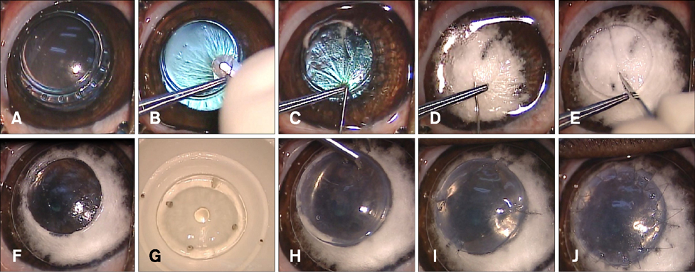
Fig. 1 Procedures for deep anterior lamellar keratoplasty using the big bubble technique. (A) The center of the cornea was trephined 750 µm using an 8 mm vacuum trephine. (B) A partial-thickness superficial anterior keratectomy was performed. (C) A 30-gauge needle attached to a 5 cc syringe was inserted at the base of the trephination gutter into the corneal stroma. (D) 4 mL of air were gently injected, causing Descemet's membrane (DM) to separate from the deep stroma. Intrastromal blanching was observed during this procedure. (E) A blanched stroma was incised with a 15° slit-knife to allow air to escape and collapse the bubble for stroma removal. (F) DM was exposed after excising the remaining stroma using corneal scissors. (G) After DM and endothelium were stripped off, the donor cornea was punched from the endothelial side using an 8.5 mm diameter Barron punch. (H) This prepared donor corneal button was fitted onto the exposed Descemet's plane of the recipient cornea. (I) Four cardinal sutures were used with 10-0 nylon at the 3, 6, 9, and 12 clock-hour positions. (J) A single running suture was performed with 16 to 18 bites using the same suture materials.
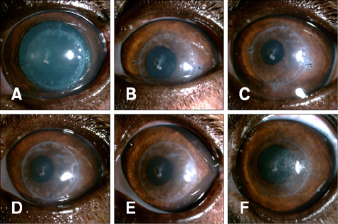
Fig. 2 Slit-lamp biomicroscopy with direct diffuse illumination of the corneas under 10× magnification. (A) 12 h after surgery, (B) 7 days, (C) 14 days, (D) 21 days, (E) 28 days and (F) 150 days after surgery.
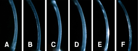
Fig. 3 Slit-lamp biomicroscopy with direct narrow slit (1 mm) under 10× magnification for the stroma. (A) 12 h after surgery, (B) 7 days, (C) 14 days, (D) 21 days, (E) 28 days and (F) 150 days after surgery.
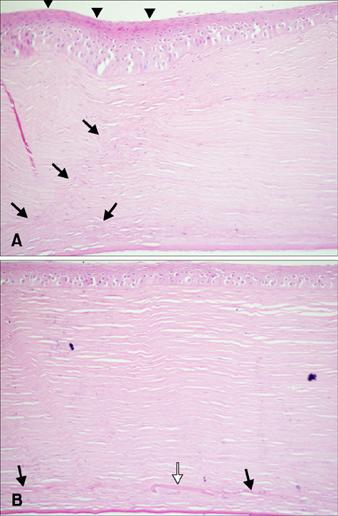
Fig. 4 Histopathological evaluation with hematoxylin and eosin staining at 150 days after surgery. (A) The epithelium was overgrown (arrowhead) at the junction of the donor and recipient corneas. In addition, deformed stromal structures were detected at the transplant junction (black arrow). (B) The central corneal part had a normal corneal structure. A small piece of DM was left in the stroma (white arrow).
Reference
-
1. Albarez de Toledo J, de la Paz MF. Manual to laser trephination in corneal transplantation: are patients noticing a difference? Br J Ophthalmol. 2014; 98:717–718.
Article2. Alio JL, Rodriguez AE, Martinez LM. Bovine pericardium membrane (tutopatch) combined with solid platelet-rich plasma for the management of perforated corneal ulcers. Cornea. 2013; 32:619–624.
Article3. Andrade AL, Laus JL, Figueiredo F, Batista CM. The use of preserved equine renal capsule to repair lamellar corneal lesions in normal dog. Vet Ophthalmol. 1999; 2:79–82.
Article4. Anwar M, Teichmann KD. Big-bubble technique to bare Descemet's membrane in anterior lamellar keratoplasty. J Cataract Refract Surg. 2002; 28:398–403.
Article5. Arenas E, Esquenazi S, Anwer M, Terry M. Lamellar corneal transplantation. Surv Ophthalmol. 2012; 57:510–529.
Article6. Bahar I, Kaiserman I, Srinivasan S, Ya-Ping J, Slomovic AR, Rootman DS. Comparison of three different techniques of corneal transplantation for keratoconus. Am J Ophthalmol. 2008; 146:905–912.e.1.
Article7. Barros PS, Garcia JA, Laus JL, Ferreira AL, Salles Gomes TL. The use of xenologous amniotic membrane to repair canine corneal perforation created by penetrating keratectomy. Vet Ophthalmol. 1998; 1:119–123.
Article8. Brightman AH, McLaughlin SA, Brogdon JD. Autogenous lamellar corneal grafting in dogs. J Am Vet Med Assoc. 1989; 195:469–475.9. Brooks DE. Penetrating keratoplasty, deep lamellar endothelial keratoplasty, and posterior lamellar keratoplasty in the horse. Clin Tech Equine Pract. 2005; 4:37–49.
Article10. Brooks DE, Plummer CE, Kallberg ME, Barrie KP, Ollivier FJ, Hendrix DV, Baker A, Scotty NC, Utter ME, Blackwood SE, Nunnery CM, Ben-Shlomo G, Gelatt KN. Corneal transplantation for inflammatory keratopathies in the horse: visual outcome in 206 cases (1993–2007). Vet Ophthalmol. 2008; 11:123–133.
Article11. Bussieres M, Krohne SG, Stiles J, Townsend WM. The use of porcine small intestinal submucosa for the repair of full-thickness corneal defects in dogs, cats and horses. Vet Ophthalmol. 2004; 7:352–359.
Article12. Fantes FE, Hanna KD, Waring GO 3rd, Pouliquen Y, Thompson KP, Savoldelli M. Wound healing after excimer laser keratomileusis (photorefractive keratectomy) in monkeys. Arch Ophthalmol. 1990; 108:665–675.
Article13. Fontana L, Parente G, Tassinari G. Clinical outcomes after deep anterior lamellar keratoplasty using the big-bubble technique in patients with keratoconus. Am J Ophthalmol. 2007; 143:117–124.
Article14. Karimian F, Feizi S. Deep anterior lamellar keratoplasty: indications, surgical techniques and complications. Middle East Afr J Ophthalmol. 2010; 17:28–37.15. Lang GK, Green WR, Maumenee AE. Clinicopathologic studies of keratoplasty eyes obtained post mortem. Am J Ophthalmol. 1986; 101:28–40.
Article16. Leiva M, Costa D, Naranjo C, Peña MT. Big bubble and intrastromal air injection techniques for performing deep anterior lamellar keratectomy in the dog. In : 2013 ECVO Annual Conference: the Congress of the European College of Veterinary Ophthalmologists; 16-19 May 2013; Barcelona, Spain.17. Levinger E, Trivzki O, Levinger S, Kremer I. Outcome of "mushroom" pattern femtosecond laser-assisted keratoplasty versus conventional penetrating keratoplasty in patients with keratoconus. Cornea. 2014; 33:481–485.18. Martins BC, Brooks DE, Plummer CE, Samuelson DA, Mangan BG, Laus JL. Light microscopic evaluation and scanning electron microscopic analysis of horse eyes following deep anterior lamellar keratectomy. Vet Ophthalmol. 2013; 16:Suppl 1. 42–51.
Article19. Melles GR, Lander F, Rietveld FJ, Remeijer L, Beekhuis WH, Binder PS. A new surgical technique for deep stromal, anterior lamellar keratoplasty. Br J Ophthalmol. 1999; 83:327–333.
Article20. McEntyre JM. Experimental penetrating keratoplasty in the dog. Arch Ophthalmol. 1968; 80:372–376.
Article21. McKee HD, Irion LC, Carley FM, Jhanji V, Brahma AK. Dissection plane of the clear margin big-bubble in deep anterior lamellar keratoplasty. Cornea. 2013; 32:e51–e52.
Article22. Niederer RL, Perumal D, Sherwin T, McGhee CN. Corneal innervation and cellular changes after corneal transplantation: an in vivo confocal microscopy study. Invest Ophthalmol Vis Sci. 2007; 48:621–626.
Article23. Panda A, Vanathi M, Kumar A, Dash Y, Priya S. Corneal graft rejection. Surv Ophthalmol. 2007; 52:375–396.
Article24. Parshall CJ Jr. Lamellar corneal-scleral transposition. J Am Anim Hosp Assoc. 1973; 9:270–277.25. Soontornvipart K, Tuntivanich N, Kecova H, Raušer P. Conjunctival pedicle graft in dogs and cats: a retrospective study of 88 cases. Acta Vet Brno. 2003; 72:63–69.
Article26. Sugita J, Kondo J. Deep lamellar keratoplasty with complete removal of pathological stroma for vision improvement. Br J Ophthalmol. 1997; 81:184–188.
Article27. Tham VM, Abbott RL. Corneal graft rejection: recent updates. Int Ophthalmol Clin. 2002; 42:105–113.
Article28. Tsubota K, Kaido M, Monden Y, Satake Y, Bissen-Miyajima H, Shimazaki J. A new surgical technique for deep lamellar keratoplasty with single running suture adjustment. Am J Ophthalmol. 1998; 126:1–8.
Article
- Full Text Links
-
- Actions
-
Cited
- CITED
-
- Close
- Share
-
- Similar articles
-
- Comparison of Early Clinical Result of Deep Anterior Lamellar Keratoplasty Using FSlaser Versus Manual Trephine
- Three Cases of Urrets-Zavalia Syndrome Following Deep Lamellar Keratoplasty (DLKP)
- A Case of Double Descemet's Membrane after Penetrating Keratoplasty Converted from Deep Anterior Lamellar Keratoplasty
- Clinical Evaluation of Full-thickness Deep Lamellar Keratoplasty
- Comparison of Deep Anterior Lamellar Keratoplasty and Penetrating Keratoplasty for Keratoconus
