Effect of Cataract Grade according to Wide-Field Fundus Images on Measurement of Macular Thickness in Cataract Patients
- Affiliations
-
- 1Department of Ophthalmology, Korea University College of Medicine, Seoul, Korea. hippotate@hanmail.net
- KMID: 2412795
- DOI: http://doi.org/10.3341/kjo.2017.0067
Abstract
- PURPOSE
To investigate the effects of cataract grade based on wide-field fundus imaging on macular thickness measured by spectral domain optical coherence tomography (SD-OCT) and its signal-to-noise ratio (SNR).
METHODS
Two hundred cataract patients (200 eyes) with preoperative measurements by wide-field fundus imaging and macular SD-OCT were enrolled. Cataract severity was graded from 1 to 4 according to the degree of macular obscuring by cataract artifact in fundus photo images. Cataract grade based on wide-field fundus image, the Lens Opacity Classification System III, macular thickness, and SD-OCT SNR were compared. All SD-OCT B-scan images were evaluated to detect errors in retinal layer segmentation.
RESULTS
Cataract grade based on wide-field fundus imaging was positively correlated with grade of posterior subcapsular cataracts (rho = 0.486, p < 0.001), but not with nuclear opalescence or cortical cataract using the Lens Opacity Classification System III. Cataract grade was negatively correlated with total macular thickness (rho = −0.509, p < 0.001) and SD-OCT SNR (rho = −0.568, p < 0.001). SD-OCT SNR was positively correlated with total macular thickness (rho = 0.571, p < 0.001). Of 200 eyes, 97 (48.5%) had segmentation errors on SD-OCT. As cataract grade increased and SD-OCT SNR decreased, the percentage of eyes with segmentation errors on SD-OCT increased. All measurements of macular thickness in eyes without segmentation errors were significantly greater than those of eyes with segmentation errors.
CONCLUSIONS
Posterior subcapsular cataracts had profound effects on cataract grade based on wide-field fundus imaging. As cataract grade based on wide-field fundus image increased, macular thickness tended to be underestimated due to segmentation errors in SD-OCT images. Segmentation errors in SD-OCT should be considered when evaluating macular thickness in eyes with cataracts.
Keyword
MeSH Terms
Figure
-

Fig. 1 Cataract grade according to standard photos of wide-field fundus imaging. (A) Grade 1, clearly visible or slightly obscured macular area. (B) Grade 2, mildly obscured macular area. (C) Grade 3, moderately obscured, but distinguishable macular area. (D) Grade 4, severely obscured, undistinguishable macular area.
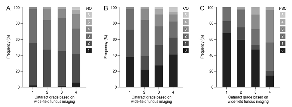
Fig. 2 Percentage constituents of nuclear opalescence (NO), cortical opacities (CO), and posterior subcapsular cataracts (PSC) with the Lens Opacities Classification System III (LOCS III) in cataract grade based on wide-field fundus imaging. (A) NO with the LOCS III. (B) CO with the LOCS III. (C) PSC with the LCOS III.

Fig. 3 Cataract grade 4 eye without segmentation errors on spectral domain optical coherence tomography. (A) Wide-field fundus imaging shows a dense localized cataract artifact. (B) Spectral domain optical coherence tomography measurements do not show segmentation errors in either the inner or outer boundary line.
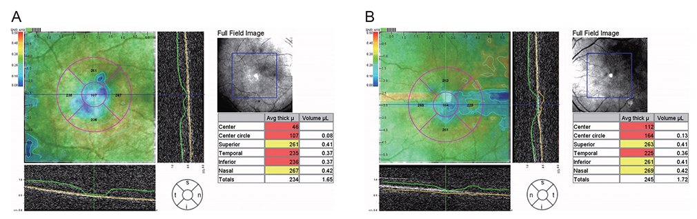
Fig. 4 Eyes with segmentation errors on spectral domain optical coherence tomography (SD-OCT). (A) A segmentation error on SD-OCT is observed only in the inner boundary line, and the automated segmentation line identifies the inner boundary line posterior to the actual inner limiting membrane. (B) A segmentation error on SD-OCT is observed in both the inner and outer boundary lines, and the automated segmentation line identifies the inner boundary line posterior to the actual inner limiting membrane and the outer boundary line posterior to the actual retinal pigment epithelium.
Reference
-
1. Fujimoto JG, Brezinski ME, Tearney GJ, et al. Optical biopsy and imaging using optical coherence tomography. Nat Med. 1995; 1:970–972.
Article2. Leung CK. Diagnosing glaucoma progression with optical coherence tomography. Curr Opin Ophthalmol. 2014; 25:104–111.
Article3. Xu G, Weinreb RN, Leung CK. Retinal nerve fiber layer progression in glaucoma: a comparison between retinal nerve fiber layer thickness and retardance. Ophthalmology. 2013; 120:2493–2500.4. Savastano MC, Minnella AM, Tamburrino A, et al. Differential vulnerability of retinal layers to early age-related macular degeneration: evidence by SD-OCT segmentation analysis. Invest Ophthalmol Vis Sci. 2014; 55:560–566.
Article5. Belair ML, Kim SJ, Thorne JE, et al. Incidence of cystoid macular edema after cataract surgery in patients with and without uveitis using optical coherence tomography. Am J Ophthalmol. 2009; 148:128–135.6. Lee DW, Kim JM, Park KH, et al. Effect of media opacity on retinal nerve fiber layer thickness measurements by optical coherence tomography. J Ophthalmic Vis Res. 2010; 5:151–157.7. Mastropasqua L, Carpineto P, Ciancaglini M, et al. Reproducibility of nerve fiber layer thickness measurements using optical coherence tomography in silicone oil-filled eyes. Ophthalmologica. 2001; 215:91–96.
Article8. El-Ashry M, Appaswamy S, Deokule S, Pagliarini S. The effect of phacoemulsification cataract surgery on the measurement of retinal nerve fiber layer thickness using optical coherence tomography. Curr Eye Res. 2006; 31:409–413.
Article9. Kim NR, Lee H, Lee ES, et al. Influence of cataract on time domain and spectral domain optical coherence tomography retinal nerve fiber layer measurements. J Glaucoma. 2012; 21:116–122.
Article10. Kok PH, van den Berg TJ, van Dijk HW, et al. The relationship between the optical density of cataract and its influence on retinal nerve fibre layer thickness measured with spectral domain optical coherence tomography. Acta Ophthalmol. 2013; 91:418–424.
Article11. Nakatani Y, Higashide T, Ohkubo S, et al. Effect of cataract and its removal on ganglion cell complex thickness and peripapillary retinal nerve fiber layer thickness measurements by fourier-domain optical coherence tomography. J Glaucoma. 2013; 22:447–455.
Article12. von Jagow B, Ohrloff C, Kohnen T. Macular thickness after uneventful cataract surgery determined by optical coherence tomography. Graefes Arch Clin Exp Ophthalmol. 2007; 245:1765–1771.
Article13. van Velthoven ME, van der Linden MH, de Smet MD, et al. Influence of cataract on optical coherence tomography image quality and retinal thickness. Br J Ophthalmol. 2006; 90:1259–1262.14. Friberg TR, Gupta A, Yu J, et al. Ultrawide angle fluorescein angiographic imaging: a comparison to conventional digital acquisition systems. Ophthalmic Surg Lasers Imaging. 2008; 39:304–311.
Article15. Wessel MM, Aaker GD, Parlitsis G, et al. Ultra-wide-field angiography improves the detection and classification of diabetic retinopathy. Retina. 2012; 32:785–791.
Article16. Mudvari SS, Virasch VV, Singa RM, MacCumber MW. Ultra-wide-field imaging for cytomegalovirus retinitis. Ophthalmic Surg Lasers Imaging. 2010; 41:311–315.
Article17. Silva PS, Cavallerano JD, Sun JK, et al. Peripheral lesions identified by mydriatic ultrawide field imaging: distribution and potential impact on diabetic retinopathy severity. Ophthalmology. 2013; 120:2587–2595.
Article18. Inoue M, Yanagawa A, Yamane S, et al. Wide-field fundus imaging using the Optos Optomap and a disposable eyelid speculum. JAMA Ophthalmol. 2013; 131:226.
Article19. Chylack LT Jr, Wolfe JK, Singer DM, et al. The Lens Opacities Classification System III: the Longitudinal Study of Cataract Study Group. Arch Ophthalmol. 1993; 111:831–836.20. Choi H, Eom Y, Kang SY, et al. Incidence of complications in cataract surgery according to the availability of partial coherence laser interferometry. J Korean Ophthalmol Soc. 2017; 58:804–810.
Article21. Domalpally A, Danis RP, Myers D, Kruse CN. Quantitative analysis of the Stratus optical coherence tomography fast macular thickness map reports. Indian J Ophthalmol. 2010; 58:131–136.
Article22. Balasubramanian M, Bowd C, Vizzeri G, et al. Effect of image quality on tissue thickness measurements obtained with spectral domain-optical coherence tomography. Opt Express. 2009; 17:4019–4036.
Article23. Chua BE, Mitchell P, Cumming RG. Effects of cataract type and location on visual function: the Blue Mountains Eye Study. Eye (Lond). 2004; 18:765–772.
Article24. Lobo CL, Faria PM, Soares MA, et al. Macular alterations after small-incision cataract surgery. J Cataract Refract Surg. 2004; 30:752–760.
Article25. McColgin AZ, Heier JS. Control of intraocular inflammation associated with cataract surgery. Curr Opin Ophthalmol. 2000; 11:3–6.
Article26. Akcay BI, Bozkurt TK, Guney E, et al. Quantitative analysis of macular thickness following uneventful and complicated cataract surgery. Clin Ophthalmol. 2012; 6:1507–1511.27. Moghimi S, Zandian M, Latifi G, et al. Topical latanoprost does not cause macular thickening after uncomplicated cataract surgery. J Ophthalmic Vis Res. 2012; 7:289–294.28. Sari ES, Ermis SS, Yazici A, et al. The effect of intracameral anesthesia on macular thickness and ganglion cell-inner plexiform layer thickness after uneventful phacoemulsification surgery: prospective and randomized controlled trial. Graefes Arch Clin Exp Ophthalmol. 2014; 252:433–439.
Article29. Falcao MS, Goncalves NM, Freitas-Costa P, et al. Choroidal and macular thickness changes induced by cataract surgery. Clin Ophthalmol. 2014; 8:55–60.30. Cagini C, Fiore T, Iaccheri B, et al. Macular thickness measured by optical coherence tomography in a healthy population before and after uncomplicated cataract phacoemulsification surgery. Curr Eye Res. 2009; 34:1036–1041.
Article
- Full Text Links
-
- Actions
-
Cited
- CITED
-
- Close
- Share
-
- Similar articles
-
- Changes in Macular Thickness after Cataract Surgery According to Optical Coherence Tomography
- Retinal Thickness After Cataract Surgery Measured by Optical Coherence Tomography
- Evaluation of Changes of Macular Thickness in Diabetic Retinopathy after Cataract Surgery
- A Case of Secondary Macular Hole Formation after Phacoemulsification in a Vitrectomized Eye
- The Comparison of SLO Retromode Images with Conventional Fundus Photography for the Detection of Drusen
