Isolated pulmonary Langerhans cell histiocytosis in a 10-month-old infant
- Affiliations
-
- 1Department of Pediatrics, Severance Children's Hospital, Yonsei University College of Medicine, Seoul, Korea. CJ@yuhs.ac
- KMID: 2412522
- DOI: http://doi.org/10.4168/aard.2018.6.3.179
Abstract
- Langerhans cell histiocytosis (LCH) is characterized by clonal proliferation and accumulation of abnormal dendritic (Langerhans) cells in various organs. Pulmonary involvement, although rare in children, has been reported in 20%-50% of childhood cases of multisystem LCH. Isolated pulmonary LCH in children, especially in infants, is still rarer, but should be suspected in those with cystic lung disease. We report a case of a 10-month-old boy who presented with chronic dyspnea and whose chest computed tomography (CT) scan demonstrated cystic lesions. Lung biopsy established the diagnosis of LCH; microscopy revealed a background of lymphocytes and eosinophils with kidney-shaped abnormal cells. These abnormal cells were positive for S-100, CD207 (Langerin), and CD1a on immunohistochemical staining. Chemotherapy was administered using a cytotoxic agent (vinblastine) and a steroid. After 12 weeks of induction chemotherapy, although no significant change in cyst size was noted on chest CT, clinical symptoms improved. Consolidation chemotherapy was then administered for 1 year. Thereafter, chest CT findings demonstrated a significant decrease in cyst size and a significant increase in the volume of normal lung parenchyma. Therefore, aggressive treatment of isolated pulmonary LCH in infants with severe tissue destruction and symptoms seems warranted.
MeSH Terms
Figure
-
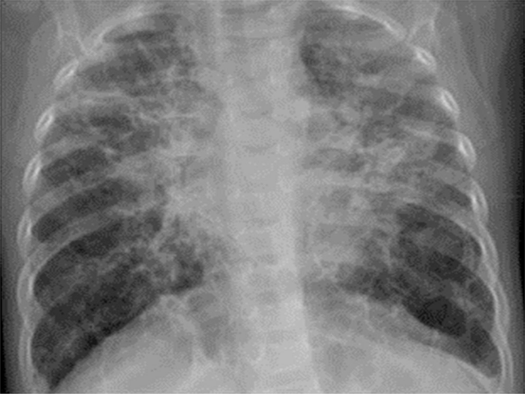
Fig. 1. A chest x-ray revealed that lung base has increased lucency due to cystic change, hyperinflated lungs with a reticular pattern.

Fig. 2. (A) A chest computed tomography (CT) at the level of the lung bases shows multiple thin-walled cysts with hardly any normal intervening lung parenchyma in the lower lobes. (B) A chest CT at the level of the upper lobes shows multiple thin-walled cyst. (C, D) A chest CT was performed after induction chemotherapy including vinblastine and steroid for response assessment. (C) A chest CT present increased size and number of multiple cystic lesions in Rt. lower lobe and decreased size in Lt. lower lobe. (D) A chest CT present increased size of cyst in both upper lung. (E, F) A chest CT was performed after consolidation chemotherapy for response assessment. The range and size of the cyst significantly decreased, and the volume of the normal lung parenchyma increased.

Fig. 3. (A) Peribronchial cellular infiltrate composed of neoplastic cells with histiocytoid features (H&E, ×100). (B) On high-power magnification the neoplastic cells are seen to display kidney shaped groove nuclear and cytoplasmic features of Langerhans cell (H&E, ×200). (C) A panel of microphotographs; CD1a cytoplasmic and membranous positivity on immunohistochemical stain (CD1a, ×200). (D) S100 positivity in the atypical histiocytes on LBC cytospin smears (S100, ×200).
Reference
-
1. Picarsic J, Jaffe R. Nosology and pathology of Langerhans cell histiocytosis. Hematol Oncol Clin North Am. 2015; 29:799–823.
Article2. Weitzman S, Egeler RM. Langerhans cell histiocytosis: update for the pe-diatrician. Curr Opin Pediatr. 2008; 20:23–9.
Article3. El Demellawy D, Young JL, de Nanassy J, Chernetsova E, Nasr A. Langerhans cell histiocytosis: a comprehensive review. Pathology. 2015; 47:294–301.
Article4. Elia D, Torre O, Cassandro R, Caminati A, Harari S. Pulmonary Langerhans cell histiocytosis: a comprehensive analysis of 40 patients and literature review. Eur J Intern Med. 2015; 26:351–6.
Article5. Odame I, Li P, Lau L, Doda W, Noseworthy M, Babyn P, et al. Pulmonary Langerhans cell histiocytosis: a variable disease in childhood. Pediatr Blood Cancer. 2006; 47:889–93.
Article6. Braier J, Latella A, Balancini B, Castaños C, Rosso D, Chantada G, et al. Outcome in children with pulmonary Langerhans cell Histiocytosis. Pediatr Blood Cancer. 2004; 43:765–9.
Article7. Sawalha L, Kumar A, Arshad A, Mador MJ. Pulmonary Langerhans cell histiocytosis: radiologic resolution following cessation of second hand smoking. Clin Respir J. 2017; 11:1063–7.
Article8. Kasmani R, Narwal-Chadha R, Naveed S, Sahoo D. Isolated pulmonary Langerhans cell histiocytosis. QJM. 2009; 102:741–2.
Article9. Kanik-Yuksek S, Ozkaya-Parlakay A, Gulhan B, Ozyoruk D, Karakus E, Cinel G, et al. A rare diagnosis in children: isolated pulmonary Langerhans cell histiocytosis. Clin Respir J. 2018; 12:355–6.
Article10. Mukhopadhyay S, Eckardt SM, Scalzetti EM. Diagnosis of pulmonary Langerhans cell histiocytosis by CT-guided core biopsy of lung: a report of three cases. Thorax. 2010; 65:833–5.
Article11. Aricò M, Girschikofsky M, Généreau T, Klersy C, McClain K, Grois N, et al. Langerhans cell histiocytosis in adults. Report from the International Registry of the Histiocyte Society. Eur J Cancer. 2003; 39:2341–8.12. Minami M, Shima T, Kato K, Yamamoto H, Tsuchihashi K, Oku S, et al. Successful treatment of adult Langerhans cell histiocytosis with intensi-fied chemotherapy. Int J Hematol. 2015; 102:244–8.
Article13. Kim HJ, Lee KS, Johkoh T, Tomiyama N, Lee HY, Han J, et al. Pulmonary Langerhans cell histiocytosis in adults: high-resolution CT-pathology comparisons and evolutional changes at CT. Eur Radiol. 2011; 21:1406–15.
Article14. Varkki S, Tergestina M, Bhonsle VS, Moses PD, Mathai J, Korula S. Isolated pulmonary Langerhans cell histiocytosis. Indian J Pediatr. 2013; 80:700–3.
Article15. Wang Q, Xia W, Zhao DY. Isolated pulmonary Langerhans cell histiocytosis in a two-year-old child: case report and literature review. Zhonghua Er Ke Za Zhi. 2012; 50:146–50.
- Full Text Links
-
- Actions
-
Cited
- CITED
-
- Close
- Share
-
- Similar articles
-
- Pulmonary Langerhans Cell Histiocytosis Accompanied by Active Pulmonary Tuberculosis
- Isolated Thymic Langerhans Cell Histiocytosis
- A Case of Pulmonary Langerhans Cell Histiocytosis with Pneumothorax
- Infant Langerhans Cell Histiocytosis Presented as Isolated Splenomegaly and Prolonged Fever
- Spontaneous Pneumothorax due to Pulmonary Invasion in Multisystemic Langerhans Cell Histiocytosis: A case report
