Arthroscopic Excision of an Intraarticular Osteoid Osteoma in the Distal Femur
- Affiliations
-
- 1Department of Orthopaedic Surgery, Dongguk University College of Medicine, Gyeongju, Korea. kjpil@dongguk.ac.kr
- KMID: 2412332
- DOI: http://doi.org/10.4055/cios.2016.8.4.475
Abstract
- An intraarticular osteoid osteoma of the knee is uncommon, and its treatment is challenging. The authors present a case of arthroscopic excision of an intraarticular osteoid osteoma in the distal femur, which was accessible through the knee joint. After confirming the nidus of the osteoid osteoma by computed tomography, the lesion was completely removed arthroscopically. The patient reported complete pain relief immediately after surgery. This case demonstrates that intraarticular osteoid osteomas in the knee joint can be treated by arthroscopic excision and that good results can be obtained.
Keyword
MeSH Terms
Figure
-

Fig. 1 (A) Sagittal T1-weighted magnetic resonance imaging (MRI) showing a 6 mm × 3.5 mm × 3 mm sized intermediate-low signal intensity nodular lesion. (B) Sagittal T2-weighted MRI showing a high signal intensity lesion with central intermediate-low signal intensity in the anterosuperior area of the lateral femoral condyle (arrowheads) with adjacent bone marrow edema.

Fig. 2 Axial (A) and sagittal (B) computed tomography images showing a 5 mm × 3 mm × 3 mm sized lesion with a central nidus and perinidal sclerosis in the anterolateral subcortical area of the distal femur.
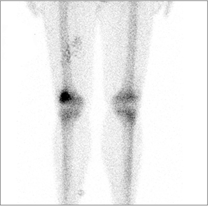
Fig. 3 Bone scan showing increased uptake in the lateral condyle of the right distal femur.

Fig. 4 (A) Arthroscopic image demonstrating a slightly elevated bony lesion at the bottom of the suprapatellar recess near the superior articular margin of the lateral femoral condyle with surrounding minimal hypertrophic synovium. (B) C-arm image taken with a 20-gauge needle held in place to confirm the correct position of the lesion. (C) Removal of the synovium over the lesion using a punching forceps and an electrocautery device. (D) Grossly reddish dense nidus of the osteoid with interconnected trabeculae observed after removal of the sclerotic rim. (E) Excision of the lesion using a small osteotome and curettes. (F) Arthroscopic confirmation of the complete removal of the reactive bone walls performed using a motorized burr.
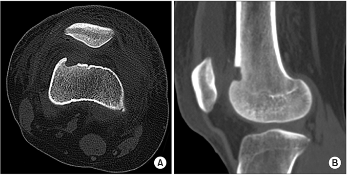
Fig. 5 Axial (A) and sagittal (B) postoperative computed tomography images showing the correct location of the removed lesion and complete nidus removal.
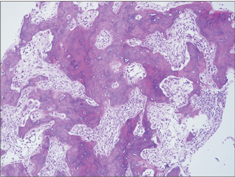
Fig. 6 Histological section of the nidus demonstrating an osteoid and woven bone with interconnected trabeculae in the background of fibrous connective tissue (H&E, × 40). These findings were used to confirm the diagnosis of osteoid osteoma.
Reference
-
1. Campanacci M, Ruggieri P, Gasbarrini A, Ferraro A, Campanacci L. Osteoid osteoma: direct visual identification and intralesional excision of the nidus with minimal removal of bone. J Bone Joint Surg Br. 1999; 81(5):814–820.2. Abnousi F, Saliman JD, Fanton GS. Arthroscopic visualization and assisted excision of osteoid osteoma at the knee: a case report and review. Am J Sports Med. 2008; 36(2):375–378.
Article3. Franceschi F, Marinozzi A, Papalia R, Longo UG, Gualdi G, Denaro E. Intra- and juxta-articular osteoid osteoma: a diagnostic challenge: misdiagnosis and successful treatment: a report of four cases. Arch Orthop Trauma Surg. 2006; 126(10):660–667.
Article4. Sluga M, Windhager R, Pfeiffer M, Dominkus M, Kotz R. Peripheral osteoid osteoma: is there still a place for traditional surgery? J Bone Joint Surg Br. 2002; 84(2):249–251.5. Heuijerjans W, Dandy DJ, Harris D. Arthroscopic excision of an intra-articular osteoid osteoma at the knee. Arthroscopy. 1986; 2(4):215–216.
Article6. Rizzello G, Longo UG, Maffulli N, Denaro V. Arthroscopic removal of an intraarticular osteoid osteoma of the distal tibia. J Foot Ankle Surg. 2010; 49(4):398.e17–398.e21.
Article7. Nehme AH, Bou Ghannam AG, Imad JP, Jabbour FC, Moucharafieh R, Wehbe J. Arthroscopic excision of intraarticular hip osteoid osteoma: a report of 2 cases. Case Rep Orthop. 2012; 2012:820501.
Article8. Gunes T, Erdem M, Bostan B, Sen C, Sahin SA. Arthroscopic excision of the osteoid osteoma at the distal femur. Knee Surg Sports Traumatol Arthrosc. 2008; 16(1):90–93.
Article9. Kelly AM, Selby RM, Lumsden E, O'Brien SJ, Drakos MC. Arthroscopic removal of an osteoid osteoma of the shoulder. Arthroscopy. 2002; 18(7):801–806.
Article10. Nourissat G, Kakuda C, Dumontier C. Arthroscopic excision of osteoid osteoma of the elbow. Arthroscopy. 2007; 23(7):799.e1–799.e4.
Article
- Full Text Links
-
- Actions
-
Cited
- CITED
-
- Close
- Share
-
- Similar articles
-
- Osteoid Osteoma of the patella: a case report
- Arthroscopic Treatment of an Osteoid Osteoma in the Femoral Neck: A Case Report
- Percutaneous Ablation of Osteoid Osteoma under Image Intensifier Guidance: A Case Report
- Arthroscopic Excision of Delayed Diagnosed Intra-articular Osteoid Osteoma of the Elbow: A Case Report
- Osteoid Osteoma in Intertrochanteric Cancellous Portion of the Femur in Adult: a case report
