Clin Orthop Surg.
2017 Jun;9(2):218-222. 10.4055/cios.2017.9.2.218.
Acromion Index in Korean Population and Its Relationship with Rotator Cuff Tears
- Affiliations
-
- 1Department of Orthopaedic Surgery, Samsung Medical Center, Sungkyunkwan University School of Medicine, Seoul, Korea. shoulderyoo@gmail.com
- KMID: 2412292
- DOI: http://doi.org/10.4055/cios.2017.9.2.218
Abstract
- BACKGROUND
Among the many causes of rotator cuff tears, scapular morphology is associated with the accelerating degenerative process of the rotator cuff. Acromion index (AI) was previously introduced and compared in two populations.
METHODS
We enrolled 100 Korean patients diagnosed with full-thickness rotator cuff tears by magnetic resonance imaging and intraoperative arthroscopic findings between January and December 2013. Another 100 Korean patients with an intact rotator cuff tendon identified on magnetic resonance imaging and other shoulder diseases, such as frozen shoulder and instability, were enrolled as controls. We retrospectively compared these 100 rotator cuff tear patients (mean age, 63 years) and 100 controls (mean age, 51 years) in this study. Two independent orthopedic surgeons assessed the AI on radiographs. We performed an interobserver reliability test of the AI assessment, and then compared the AI between two groups.
RESULTS
The measurement of the AI showed excellent reliability (intraclass correlation coefficient, 0.82). The mean AI in the rotator cuff tear group was 0.68 and it was significantly different between groups (p<0.001, 95% confidence interval). The AI was not related to tear size.
CONCLUSIONS
Our study showed that the AI was an effective predictive factor for rotator cuff tears in a Korean population.
Keyword
MeSH Terms
-
*Acromion/anatomy & histology/diagnostic imaging
Adult
Aged
Aged, 80 and over
Asian Continental Ancestry Group/*statistics & numerical data
Female
Humans
Magnetic Resonance Imaging
Male
Middle Aged
Prognosis
ROC Curve
Republic of Korea/epidemiology
Retrospective Studies
*Rotator Cuff Injuries/diagnostic imaging/epidemiology
Figure
-
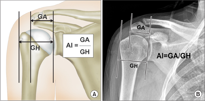
Fig. 1 Acrominon index (AI). (A) AI was defined as the ratio of the distance from the glenoid to the lateral margin of the acromion (GA) divided by the distance from the glenoid rim to the lateral aspect of the humeral head (GH). (B) Anteroposterior radiographic view of the right shoulder: calculation of AI was done according to the method described in Fig. 1A.
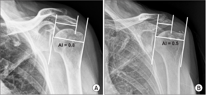
Fig. 2 Comparison of acromion index (AI) on true anteroposterior (AP) radiographs: the AI was higher in the shoulder with a rotator cuff tear (A) than in the shoulder with an intact rotator cuff (B).
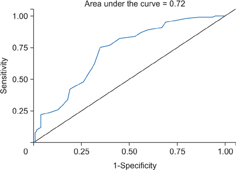
Fig. 3 Receiver operating characteristic curve for the acromion index in blue line. The reference line is indicated in black line.
Reference
-
1. Neer CS 2nd. Anterior acromioplasty for the chronic impingement syndrome in the shoulder: a preliminary report. J Bone Joint Surg Am. 1972; 54(1):41–50.
Article2. Neer CS 2nd. Impingement lesions. Clin Orthop Relat Res. 1983; (173):70–77.
Article3. Codman EA. The shoulder: rupture of the supraspinatus tendon and other lesions in or about the subacromial bursa. Boston, MA: Thomas Todd;1934.4. Bigliani LU, Morrison DS, April EW. The morphology of the acromion and its relationship to rotator cuff tears. Orthop Trans. 1986; 10:228.5. Nyffeler RW, Werner CM, Sukthankar A, Schmid MR, Gerber C. Association of a large lateral extension of the acromion with rotator cuff tears. J Bone Joint Surg Am. 2006; 88(4):800–805.
Article6. Miyazaki AN, Itoi E, Sano H, et al. Comparison between the acromion index and rotator cuff tears in the Brazilian and Japanese populations. J Shoulder Elbow Surg. 2011; 20(7):1082–1086.
Article7. DeOrio JK, Cofield RH. Results of a second attempt at surgical repair of a failed initial rotator-cuff repair. J Bone Joint Surg Am. 1984; 66(4):563–567.
Article8. Youden WJ. Index for rating diagnostic tests. Cancer. 1950; 3(1):32–35.
Article9. Kim JR, Ryu KJ, Hong IT, Kim BK, Kim JH. Can a high acromion index predict rotator cuff tears? Int Orthop. 2012; 36(5):1019–1024.
Article10. Moor BK, Wieser K, Slankamenac K, Gerber C, Bouaicha S. Relationship of individual scapular anatomy and degenerative rotator cuff tears. J Shoulder Elbow Surg. 2014; 23(4):536–541.
Article11. Torrens C, Lopez JM, Puente I, Caceres E. The influence of the acromial coverage index in rotator cuff tears. J Shoulder Elbow Surg. 2007; 16(3):347–351.
Article12. Zumstein MA, Jost B, Hempel J, Hodler J, Gerber C. The clinical and structural long-term results of open repair of massive tears of the rotator cuff. J Bone Joint Surg Am. 2008; 90(11):2423–2431.
Article13. Hamid N, Omid R, Yamaguchi K, Steger-May K, Stobbs G, Keener JD. Relationship of radiographic acromial characteristics and rotator cuff disease: a prospective investigation of clinical, radiographic, and sonographic findings. J Shoulder Elbow Surg. 2012; 21(10):1289–1298.
Article14. Balke M, Liem D, Greshake O, Hoeher J, Bouillon B, Banerjee M. Differences in acromial morphology of shoulders in patients with degenerative and traumatic supraspinatus tendon tears. Knee Surg Sports Traumatol Arthrosc. 2016; 24(7):2200–2205.
Article15. Ames JB, Horan MP, Van der Meijden OA, Leake MJ, Millett PJ. Association between acromial index and outcomes following arthroscopic repair of full-thickness rotator cuff tears. J Bone Joint Surg Am. 2012; 94(20):1862–1869.
Article16. Fujisawa Y, Mihata T, Murase T, Sugamoto K, Neo M. Three-dimensional analysis of acromial morphologic characteristics in patients with and without rotator cuff tears using a reconstructed computed tomography model. Am J Sports Med. 2014; 42(11):2621–2626.
Article17. Matthews TJ, Hand GC, Rees JL, Athanasou NA, Carr AJ. Pathology of the torn rotator cuff tendon: reduction in potential for repair as tear size increases. J Bone Joint Surg Br. 2006; 88(4):489–495.18. Bigliani LU, Ticker JB, Flatow EL, Soslowsky LJ, Mow VC. The relationship of acromial architecture to rotator cuff disease. Clin Sports Med. 1991; 10(4):823–838.
Article19. Walch G, Liotard JP, Boileau P, Noel E. Postero-superior glenoid impingement: another shoulder impingement. Rev Chir Orthop Reparatrice Appar Mot. 1991; 77(8):571–574.20. Sher JS, Uribe JW, Posada A, Murphy BJ, Zlatkin MB. Abnormal findings on magnetic resonance images of asymptomatic shoulders. J Bone Joint Surg Am. 1995; 77(1):10–15.
Article
- Full Text Links
-
- Actions
-
Cited
- CITED
-
- Close
- Share
-
- Similar articles
-
- Factors Influencing the Restoration of Acromiohumeral Distance of Immediate Postoperative Period in Patients Who Have Rotator Cuff Repair Surgery with Large-to-Massive Rotator Cuff Tears
- Partial-thickness rotator cuff tears: a review of current literature on evaluation and management
- The Effect of the Acromion Shape on Rotator Cuff Tears
- A Retrospective Analysis of the Relationship Between Rotator Cuff Tear and Biceps Lesion
- Correlation Between Degree of Torn Rotator Cuff in MRI and Degenerative Change of Acromion and Greater Tuberosity in Simple Radiography
