Clin Orthop Surg.
2017 Dec;9(4):397-404. 10.4055/cios.2017.9.4.397.
Long-term Results of Modified Salter Innominate Osteotomy for Legg-Calvé-Perthes Disease
- Affiliations
-
- 1Department of Orthopaedic Surgery, Center for Joint Disease, Chonnam National University Hwasun Hospital, Hwasun, Korea. tryoon@jnu.ac.kr
- 2Department of Traumatology, Neurosurgery, and Military Field Surgery, Samarkand State Medical Institute, Samarkand, Uzbekistan.
- KMID: 2412239
- DOI: http://doi.org/10.4055/cios.2017.9.4.397
Abstract
- BACKGROUND
In a previous study, we reported clinical and radiographic results of our modified Salter innominate osteotomy technique in 16 hips affected by Legg-Calvé-Perthes disease (LCPD) with an average follow-up of 31.8 months. In this study, we present the long-term results of the osteotomy in LCPD patients followed until physeal closure.
METHODS
Thirty hips of 29 patients were followed until skeletal maturation after modified Salter innominate osteotomy. The mean follow-up duration was 12.9 years (range, 9.1 to 16.0 years). Eleven hips (36.7%) were classified as Catterall group III and 19 (63.3%) as Catterall group VI. Stable interposition of a bone block was achieved using one biodegradable screw in nine hips and without any fixation device in 21 hips by simply changing the direction of osteotomy. The Harris Hip Score (HHS), Western Ontario and McMaster Universities Osteoarthritis Index (WOMAC) score, and hip function were evaluated at the last follow-up. Radiological outcomes were evaluated using Wiberg's center-edge angle, the Mose method, and Stulberg classification, and osteoarthritic changes were evaluated using the Tonnis classification.
RESULTS
Mean HHS and WOMAC score were 80.2 points and 54 points, respectively, preoperatively and these were improved to 96.2 points and 28 points, respectively, at the last follow-up. Clinical results, according to Robinson's criteria, were good in 18, fair in seven, and poor in five hips. Radiological results assessed using the Mose method were good in 18, fair in six, and poor in six hips, and according to the Stulberg classification, nine hips were class I, nine were class II, eight were class III, and four were class IV. The mean center-edge angle improved from 19.7° preoperatively to 29.6° at the final follow-up. According the Tonnis classification, three hips were grade 2, five were grade 1, and 22 were grade 0. Of the three grade 2 hips, two underwent Chiari osteotomy 12.1 and 8.8 years postoperatively, and the other underwent total hip arthroplasty 12.9 years postoperatively.
CONCLUSIONS
The modified Salter innominate osteotomy produced relatively satisfactory long-term clinical and radiological results.
MeSH Terms
-
Bone Development
Child
Child, Preschool
Female
Follow-Up Studies
Hip Joint/diagnostic imaging/*physiopathology
Humans
Legg-Calve-Perthes Disease/complications/physiopathology/*surgery
Male
Osteoarthritis, Hip/diagnostic imaging/etiology
Osteotomy/*methods
Radiography
Range of Motion, Articular
Time Factors
Treatment Outcome
Figure
-
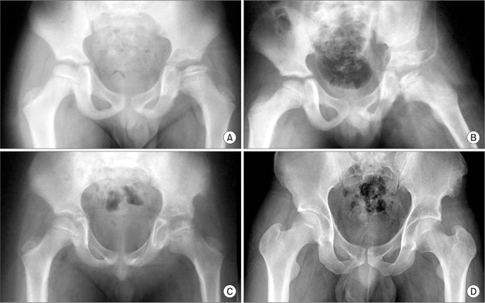
Fig. 1 Case number 22. (A) Preoperative radiograph of an 8.5-year-old male showing Legg-Calvé-Perthes disease with a Wiberg's center-edge angle of 14°. (B) Radiograph taken immediately after surgery. (C) Radiograph taken 2 years after surgery. (D) Radiograph taken 15.9 years after surgery, showing good remodeling of the acetabular osteotomy site and good femoral head sphericity.
Reference
-
1. Salter RB. Treatment by innominate osteotomy. Instr Course Lect. 1973; 22:309–316.2. Catterall A. The natural history of Perthes' disease. J Bone Joint Surg Br. 1971; 53(1):37–53. PMID: 5578764.
Article3. Millis MB, Hall JE. Transiliac lengthening of the lower extremity: a modified innominate osteotomy for the treatment of postural imbalance. J Bone Joint Surg Am. 1979; 61(8):1182–1194. PMID: 389929.
Article4. Salter RB. Role of innominate osteotomy in the treatment of congenital dislocation and subluxation of the hip in the older child. J Bone Joint Surg Am. 1966; 48(7):1413–1439. PMID: 5921797.
Article5. Yoon TR, Rowe SM, Chung JY, Song EK, Mulyadi D, Anwar IB. A new innominate osteotomy in Perthes' disease. J Pediatr Orthop. 2003; 23(3):363–367. PMID: 12724601.
Article6. Herring JA, Neustadt JB, Williams JJ, Early JS, Browne RH. The lateral pillar classification of Legg-Calve-Perthes disease. J Pediatr Orthop. 1992; 12(2):143–150. PMID: 1552014.
Article7. Harris WH. Traumatic arthritis of the hip after dislocation and acetabular fractures: treatment by mold arthroplasty. An end-result study using a new method of result evaluation. J Bone Joint Surg Am. 1969; 51(4):737–755. PMID: 5783851.8. Bellamy N, Buchanan WW, Goldsmith CH, Campbell J, Stitt LW. Validation study of WOMAC: a health status instrument for measuring clinically important patient relevant outcomes to antirheumatic drug therapy in patients with osteoarthritis of the hip or knee. J Rheumatol. 1988; 15(12):1833–1840. PMID: 3068365.9. Robinson HJ Jr, Putter H, Sigmond MB, O'Connor S, Murray KR. Innominate osteotomy in Perthes disease. J Pediatr Orthop. 1988; 8(4):426–435. PMID: 3392195.
Article10. Kitakoji T, Hattori T, Kitoh H, Katoh M, Ishiguro N. Which is a better method for Perthes' disease: femoral varus or Salter osteotomy? Clin Orthop Relat Res. 2005; (430):163–170.11. Wiberg G. Studies on dysplastic acetabula and congenital subluxation of the hip joint: with special reference to the complication of osteoarthritis. Acta Chir Scand. 1939; 83(Suppl 58):53–68.12. Mose K. Methods of measuring in Legg-Calve-Perthes disease with special regard to the prognosis. Clin Orthop Relat Res. 1980; (150):103–109. PMID: 7428206.13. Stulberg SD, Cooperman DR, Wallensten R. The natural history of Legg-Calve-Perthes disease. J Bone Joint Surg Am. 1981; 63(7):1095–1108. PMID: 7276045.14. Tonnis D. Normal values of the hip joint for the evaluation of X-rays in children and adults. Clin Orthop Relat Res. 1976; (119):39–47.15. Ingman AM, Paterson DC, Sutherland AD. A comparison between innominate osteotomy and hip spica in the treatment of Legg-Perthes' disease. Clin Orthop Relat Res. 1982; (163):141–147.
Article16. Salter RB. The present status of surgical treatment for Legg-Perthes disease. J Bone Joint Surg Am. 1984; 66(6):961–966. PMID: 6736099.
Article17. Olney BW, Asher MA. Combined innominate and femoral osteotomy for the treatment of severe Legg-Calve-Perthes disease. J Pediatr Orthop. 1985; 5(6):645–651. PMID: 4066935.18. Canale ST, D'Anca AF, Cotler JM, Snedden HE. Innominate osteotomy in Legg-Calve-Perthes disease. J Bone Joint Surg Am. 1972; 54(1):25–40. PMID: 4559945.19. Moberg A, Hansson G, Kaniklides C. Results after femoral and innominate osteotomy in Legg-Calve-Perthes disease. Clin Orthop Relat Res. 1997; (334):257–264.20. Ishida A, Kuwajima SS, Laredo Filho J, Milani C. Salter innominate osteotomy in the treatment of severe Legg-Calve-Perthes disease: clinical and radiographic results in 32 patients (37 hips) at skeletal maturity. J Pediatr Orthop. 2004; 24(3):257–264. PMID: 15105719.21. Sponseller PD, Desai SS, Millis MB. Comparison of femoral and innominate osteotomies for the treatment of Legg-Calve-Perthes disease. J Bone Joint Surg Am. 1988; 70(8):1131–1139. PMID: 3417698.22. Castaneda P, Vidal-Ruiz C, Mendez A, Salazar DP, Torres A. How often does femoroacetabular impingement occur after an innominate osteotomy for acetabular dysplasia? Clin Orthop Relat Res. 2016; 474(5):1209–1215. PMID: 26822844.23. Robb CA, Datta A, Nayeemuddin M, Bache CE. Assessment of acetabular retroversion following long term review of Salter's osteotomy. Hip Int. 2009; 19(1):8–12. PMID: 19455495.
Article24. Nakashima Y, Kubota H, Yamamoto T, Mawatari T, Motomura G, Iwamoto Y. Transtrochanteric rotational osteotomy for late-onset Legg-Calve-Perthes disease. J Pediatr Orthop. 2011; 31(2 Suppl):S223–S228. PMID: 21857443.
Article25. Wenger DR, Pandya NK. Advanced containment methods for the treatment of Perthes disease: Salter plus varus osteotomy and triple pelvic osteotomy. J Pediatr Orthop. 2011; 31(2 Suppl):S198–S205. PMID: 21857439.
- Full Text Links
-
- Actions
-
Cited
- CITED
-
- Close
- Share
-
- Similar articles
-
- Femoral Varus Osteotomy Versus Salter Innominate Osteotomy in the Treatment of Legg - Calve - Perthes Disease
- Innominate Osteotomy in Legg-Calve-Perthes Disease
- Innominate osteotomy for the treatment of Legg-Calve-Perthes disease
- A New Innominate Osteotomy in Legg-Calve-Perthes' Disease
- Subtrochanteric Varization Osteotomy with Open Wedge Technic in a Legg-Calve-Perthes Disease
