Impact of Fat Infiltration in Cervical Extensor Muscles on Cervical Lordosis and Neck Pain: A Cross-Sectional Study
- Affiliations
-
- 1Department of Orthopaedic Surgery, Kwangju Christian Hospital, Gwangju, Korea. stemcellchoi@gmail.com
- KMID: 2411745
- DOI: http://doi.org/10.4055/cios.2018.10.2.197
Abstract
- BACKGROUND
Weakness of cervical extensor muscles causes loss of cervical lordosis, which could also cause neck pain. The aim of this study was to investigate the impact of fat infiltration in cervical extensor muscles on cervical lordosis and neck pain.
METHODS
Fifty-six patients who suffered from neck pain were included in this study. Fat infiltration in cervical extensor muscles was measured at each level of C2-3 and C6-7 using axial magnetic resonance imaging. The visual analogue scale (VAS), 12-Item Short Form Health Survey (SF-12), and Neck Disability Index (NDI) were used for clinical assessment.
RESULTS
The mean fat infiltration was 206.3 mm2 (20.3%) at C2-3 and 240.6 mm2 (19.5%) at C6-7. Fat infiltration in cervical extensor muscles was associated with high VAS scores at both levels (p = 0.047 at C2-3; p = 0.009 at C6-7). At C2-3, there was a negative correlation between fat infiltration of the cervical extensor muscles and cervical lordosis (r = −0.216; p = 0.020). At C6-7, fat infiltration in the cervical extensor muscles was closely related to NDI (p = 0.003) and SF-12 (p > 0.05). However, there was no significant correlation between cervical lordosis and clinical outcomes (VAS, p = 0.112; NDI, p = 0.087; and SF-12, p > 0.05).
CONCLUSIONS
These results suggest that fat infiltration in the upper cervical extensor muscles has relevance to the loss of cervical lordosis, whereas fat infiltration in the lower cervical extensor muscles is associated with cervical functional disability.
Keyword
MeSH Terms
Figure
-
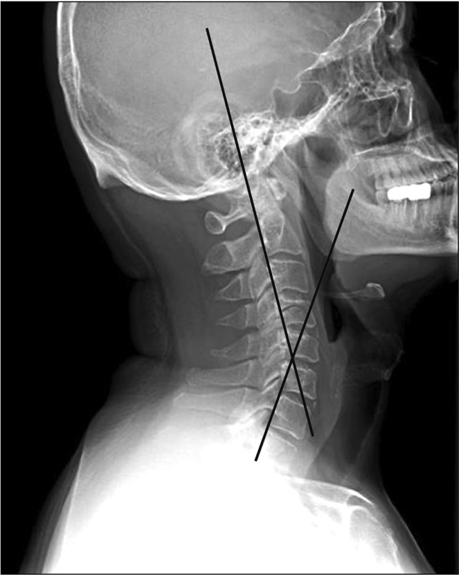
Fig. 1 Measurement of cervical lordotic angle by the posterior tangent method.
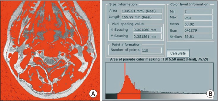
Fig. 2 Pseudo-coloring program was applied on the C2–3 axial image. (A) The muscle tissue on the magnetic resonance imaging scan is colored gray. (B) The area of pseudo-color masking of region of interest was obtained.
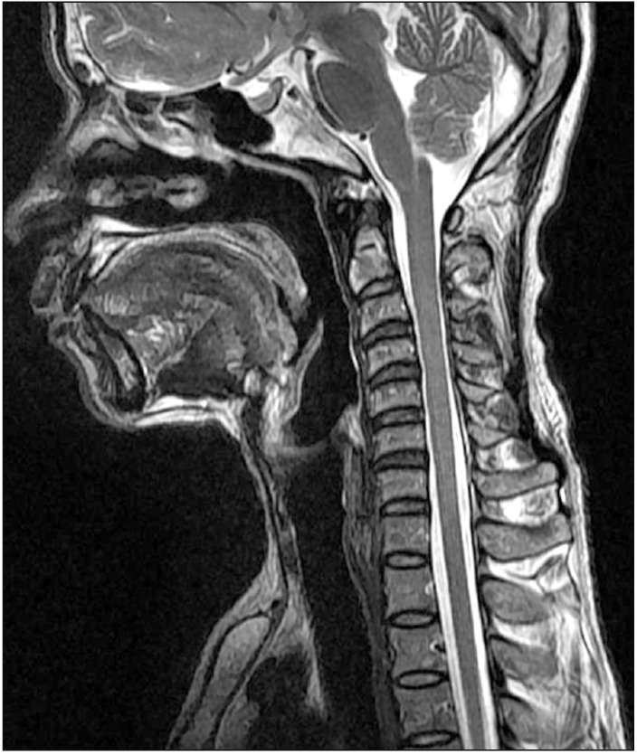
Fig. 3 Degeneration grades of the cervical intervertebral discs in magnetic resonance imaging are as follows: C2–3, grade I; C3–4, grade II; C4–5, grade II; C5–6, grade III; C6–7, grade III; and C7–T1, grade II.
Cited by 1 articles
-
Association between fatty infiltration in the cervical multifidus and treatment response following cervical interlaminar epidural steroid injection
Hyun-Jung Kwon, Chan-Sik Kim, Sungwon Kim, Syn Hae Yoon, Jungho Koh, Young Ki Kim, Seong-Soo Choi, Jin-Woo Shin, Doo-Hwan Kim
Korean J Pain. 2023;36(3):358-368. doi: 10.3344/kjp.23092.
Reference
-
1. Panjabi MM, Cholewicki J, Nibu K, Grauer J, Babat LB, Dvorak J. Critical load of the human cervical spine: an in vitro experimental study. Clin Biomech (Bristol, Avon). 1998; 13(1):11–17.
Article2. Campbell WW, Vasconcelos O, Laine FJ. Focal atrophy of the multifidus muscle in lumbosacral radiculopathy. Muscle Nerve. 1998; 21(10):1350–1353. PMID: 9736071.
Article3. Kader DF, Wardlaw D, Smith FW. Correlation between the MRI changes in the lumbar multifidus muscles and leg pain. Clin Radiol. 2000; 55(2):145–149. PMID: 10657162.
Article4. Alaranta H, Tallroth K, Soukka A, Heliovaara M. Fat content of lumbar extensor muscles and low back disability: a radiographic and clinical comparison. J Spinal Disord. 1993; 6(2):137–140. PMID: 8504225.5. van Tulder MW, Assendelft WJ, Koes BW, Bouter LM. Spinal radiographic findings and nonspecific low back pain: a systematic review of observational studies. Spine (Phila Pa 1976). 1997; 22(4):427–434. PMID: 9055372.6. Kjaer P, Bendix T, Sorensen JS, Korsholm L, Leboeuf-Yde C. Are MRI-defined fat infiltrations in the multifidus muscles associated with low back pain? BMC Med. 2007; 5:2. PMID: 17254322.
Article7. Elliott J, Sterling M, Noteboom JT, Treleaven J, Galloway G, Jull G. The clinical presentation of chronic whiplash and the relationship to findings of MRI fatty infiltrates in the cervical extensor musculature: a preliminary investigation. Eur Spine J. 2009; 18(9):1371–1378. PMID: 19672633.
Article8. Elliott J, Jull G, Noteboom JT, Darnell R, Galloway G, Gibbon WW. Fatty infiltration in the cervical extensor muscles in persistent whiplash-associated disorders: a magnetic resonance imaging analysis. Spine (Phila Pa 1976). 2006; 31(22):E847–E855. PMID: 17047533.9. Harrison DE, Harrison DD, Cailliet R, Troyanovich SJ, Janik TJ, Holland B. Cobb method or Harrison posterior tangent method: which to choose for lateral cervical radiographic analysis. Spine (Phila Pa 1976). 2000; 25(16):2072–2078. PMID: 10954638.10. Lee JC, Cha JG, Kim Y, Kim YI, Shin BJ. Quantitative analysis of back muscle degeneration in the patients with the degenerative lumbar flat back using a digital image analysis: comparison with the normal controls. Spine (Phila Pa 1976). 2008; 33(3):318–325. PMID: 18303466.11. Miyazaki M, Hong SW, Yoon SH, Morishita Y, Wang JC. Reliability of a magnetic resonance imaging-based grading system for cervical intervertebral disc degeneration. J Spinal Disord Tech. 2008; 21(4):288–292. PMID: 18525490.
Article12. Ware J Jr, Kosinski M, Keller SD. A 12-Item Short-Form Health Survey: construction of scales and preliminary tests of reliability and validity. Med Care. 1996; 34(3):220–233. PMID: 8628042.13. Vernon H, Mior S. The Neck Disability Index: a study of reliability and validity. J Manipulative Physiol Ther. 1991; 14(7):409–415. PMID: 1834753.14. McPartland JM, Brodeur RR, Hallgren RC. Chronic neck pain, standing balance, and suboccipital muscle atrophy: a pilot study. J Manipulative Physiol Ther. 1997; 20(1):24–29. PMID: 9004119.15. Anderson JS, Hsu AW, Vasavada AN. Morphology, architecture, and biomechanics of human cervical multifidus. Spine (Phila Pa 1976). 2005; 30(4):E86–E91. PMID: 15706328.
Article16. Shahidi B, Parra CL, Berry DB, et al. Contribution of lumbar spine pathology and age to paraspinal muscle size and fatty infiltration. Spine (Phila Pa 1976). 2017; 42(8):616–623. PMID: 27517512.
Article17. Shahidi B, Hubbard JC, Gibbons MC, et al. Lumbar multifidus muscle degenerates in individuals with chronic degenerative lumbar spine pathology. J Orthop Res. 2017; 35(12):2700–2706. PMID: 28480978.
Article18. Nukada H, McMorran PD, Shimizu J. Acute inflammatory demyelination in reperfusion nerve injury. Ann Neurol. 2000; 47(1):71–79. PMID: 10632103.
Article19. Andary MT, Hallgren RC, Greenman PE, Rechtien JJ. Neurogenic atrophy of suboccipital muscles after a cervical injury: a case study. Am J Phys Med Rehabil. 1998; 77(6):545–549. PMID: 9862543.20. Fleckenstein JL, Watumull D, Conner KE, et al. Denervated human skeletal muscle: MR imaging evaluation. Radiology. 1993; 187(1):213–218. PMID: 8451416.
Article21. Dulor JP, Cambon B, Vigneron P, et al. Expression of specific white adipose tissue genes in denervation-induced skeletal muscle fatty degeneration. FEBS Lett. 1998; 439(1-2):89–92. PMID: 9849884.
Article22. Maughan RJ. Relationship between muscle strength and muscle cross-sectional area: implications for training. Sports Med. 1984; 1(4):263–269. PMID: 6568749.23. Hogan KA, Manning EL, Glaser JA. Progressive cervical kyphosis associated with botulinum toxin injection. South Med J. 2006; 99(8):888–891. PMID: 16929888.
Article24. Hallgren RC, Greenman PE, Rechtien JJ. Atrophy of suboccipital muscles in patients with chronic pain: a pilot study. J Am Osteopath Assoc. 1994; 94(12):1032–1038. PMID: 7852102.25. Elliott J, Sterling M, Noteboom JT, Darnell R, Galloway G, Jull G. Fatty infiltrate in the cervical extensor muscles is not a feature of chronic, insidious-onset neck pain. Clin Radiol. 2008; 63(6):681–687. PMID: 18455560.
Article26. Harrison DE, Harrison DD, Janik TJ, William Jones E, Cailliet R, Normand M. Comparison of axial and flexural stresses in lordosis and three buckled configurations of the cervical spine. Clin Biomech (Bristol, Avon). 2001; 16(4):276–284.
Article27. Steinmetz MP, Kager CD, Benzel EC. Ventral correction of postsurgical cervical kyphosis. J Neurosurg. 2003; 98(1 Suppl):1–7.
Article
- Full Text Links
-
- Actions
-
Cited
- CITED
-
- Close
- Share
-
- Similar articles
-
- Association between fatty infiltration in the cervical multifidus and treatment response following cervical interlaminar epidural steroid injection
- Efficacy of Modified Cervical and Shoulder Retraction Exercise in Patients With Loss of Cervical Lordosis and Neck Pain
- Evaluation of the Association between Neck Pain and the Trapezius Muscles in Patients with Cervical Myelopathy Using Motor Evoked Potential: A Retrospective Study
- Radiologic Assessment of Forward Head Posture and Its Relation to Myofascial Pain Syndrome
- Forward Head Posture: Relationship between Spinal Alignment Indicies and Myoelectrical Activities of the Paraspinal Muscles
