Unintentional lumbar facet joint injection guided by fluoroscopy during interlaminar epidural steroid injection: a retrospective analysis
- Affiliations
-
- 1Department of Anesthesiology and Pain Medicine, Jeju National University School of Medicine, Jeju, Korea. solafide5@hanmail.net
- 2Department of Anesthesiology and Pain Medicine, Hankook Hospital, Korea.
- 3Jeju National University School of Medicine, Jeju, Korea.
- KMID: 2410830
- DOI: http://doi.org/10.3344/kjp.2018.31.2.87
Abstract
- BACKGROUND
An epidural steroid injection (ESI) is a commonly administered procedure in pain clinics. An unintentional lumbar facet joint injection during interlaminar ESI was reported in a previous study, but there has not been much research on the characteristics of an unintentional lumbar facet joint injection. This study illustrated the imaging features of an unintentional lumbar facet joint injection during an interlaminar ESI and analyzed characteristics of patients who underwent this injection.
METHODS
From December 2015 to May 2017, we performed 662 lumbar ESIs and we identified 24 cases (21 patients) that underwent a lumbar facet joint injection. We gathered data contrast pattern, needle approach levels and directions, injected facet joint levels and directions, presence of lumbar spine disease as seen on magnetic resonance images (MRI), and histories of lumbar spine surgeries.
RESULTS
The contrast pattern in the facet joint has a sigmoid or ovoid contrast pattern confined to the vicinity of the facet joint. The incidence of unintentional lumbar facet joint injection was 3.6%. The mean age was 68.47 years. Among these 21 patients, 14 (66.7%) were injected in the facet joint ipsilaterally to the needle approach. Among the 20 patients who received MRI, all (100%) had central stenosis and 15 patients (75%) had severe stenosis.
CONCLUSIONS
When the operator performs an interlaminar ESI on patients with central spinal stenosis, the contrast pattern on the fluoroscopy during interlaminar ESI should be carefully examined to distinguish between the epidural space and facet joint.
Keyword
MeSH Terms
Figure
-
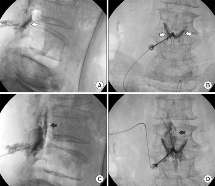
Fig. 1 Unintentional facet joint injection during interlaminar epidural steroid injection (ESI) at L4–L5. A 64-year-old woman with central and foraminal stenosis and herniated intervertebral disc (HIVD) at L4–L5 as seen on magnetic resonance image (MRI). (A) The lateral view demonstrates the contrast pattern injected on the facet joint (white arrow). (B) The anteroposterior (AP) view demonstrates the butterfly-shaped contrast pattern on both facet joints (white arrow). (C) The operator placed the needle deeper into the posterior epidural space, and the lateral view demonstrates the contrast pattern injected on the posterior epidural space (black arrow). (D) The AP view also demonstrates the contrast pattern on the epidural space (black arrow).
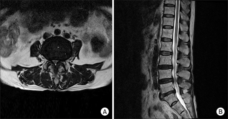
Fig. 2 MRI images of patient who underwent an unintentional facet joint injection during interlaminar ESI at L4–L5. (A) Axial T2 image. (B) Sagittal T2 image. Both images show severe central and left mild foraminal stenosis and HIVD at L4–L5.
Reference
-
1. Hong JH, Park EK, Park KB, Park JH, Jung SW. Comparison of clinical efficacy in epidural steroid injections through transforaminal or parasagittal approaches. Korean J Pain. 2017; 30(3):220–228. PMID: 28757923.
Article2. Huang AJ, Rosenthal DI, Palmer WE. Inadvertent intra-articular lumbar facet joint injection during fluoroscopically guided interlaminar epidural steroid injection. Skeletal Radiol. 2011; 40(1):33–45. PMID: 20820772.
Article3. Huang AJ, Palmer WE. Incidence of inadvertent intraarticular lumbar facet joint injection during fluoroscopically guided interlaminar epidural steroid injection. Skeletal Radiol. 2012; 41(2):157–162. PMID: 22159989.
Article4. Lehman VT, Murthy NS, Diehn FE, Verdoorn JT, Maus TP. The posterior ligamentous complex inflammatory syndrome: spread of fluid and inflammation in the retrodural space of Okada. Clin Radiol. 2015; 70(5):528–535. PMID: 25577652.
Article5. Okada K. Studies on the cervical facet joints using arthrography of the cervical facet joint (author's transl). Nihon Seikeigeka Gakkai Zasshi. 1981; 55(6):563–580. PMID: 7310204.6. Sarazin L, Chevrot A, Pessis E, Minoui A, Drape JL, Chemla N, et al. Lumbar facet joint arthrography with the posterior approach. Radiographics. 1999; 19(1):93–104. PMID: 9925394.
Article7. Murthy NS, Maus TP, Aprill C. The retrodural space of Okada. AJR Am J Roentgenol. 2011; 196(6):784–789.
Article8. McCormick CC, Taylor JR, Twomey LT. Facet joint arthrography in lumbar spondylolysis: anatomic basis for spread of contrast medium. Radiology. 1989; 171(1):193–196. PMID: 2928525.
Article9. Fenton DS, Czervionke LF. Image-guided spine intervention. Philadelphia (PA): Saunders;2003.
- Full Text Links
-
- Actions
-
Cited
- CITED
-
- Close
- Share
-
- Similar articles
-
- Spinal interventions under ultrasound guidance
- Short-Term Outcome of Fluoroscopic-Guided Steroid Injection Therapy of Lumbar Facet Cyst-Induced Radicular Pain
- Fluoroscopy-Guided Intra-Articular Facet Joint Steroid Injection for the Management of Low Back Pain: Therapeutic Effectiveness and Arthrographic Pattern
- Oblique interlaminar lumbar epidural steroid injection for management of low back pain with lumbosacral radicular pain: A case report
- Efficacy of FluoroscopyGuided Lumbar Facet Joint Synovial Cyst Rupture with Intra-Articular Steroid Injection after Laminectomy
