Falcine Sinus: Incidence and Imaging Characteristics of Three-Dimensional Contrast-Enhanced Thin-Section Magnetic Resonance Imaging
- Affiliations
-
- 1Department of Diagnostic Radiology, The First Affiliated Hospital of Sun Yat-Sen University, Guangzhou 510080, China. usefulkey0077@163.com
- 2Department of Ultrasonography, The First Affiliated Hospital of Sun Yat-Sen University, Guangzhou 510080, China.
- KMID: 2410817
- DOI: http://doi.org/10.3348/kjr.2018.19.3.463
Abstract
OBJECTIVE
To evaluate the incidence, characteristics, and variations of the falcine sinus with contrast-enhanced three-dimentional (3D) thin-section magnetic resonance (MR) images.
MATERIALS AND METHODS
retrospective review identified 1531 patients (745 males and 786 females, 2 months to 85 years) who underwent cranial MR imaging including T1-weighted imaging, T2-weighted imaging, T2-weighted fluid-attenuated inversion recovery, contrast-enhanced 3D thin-section sagittal scans, and MR venography, from June 2014 to January 2016. The incidence, characteristics of the falcine sinus, and coexisted intracranial lesions were confirmed by two neuroradiologists.
RESULTS
Falcine sinuses were identified in 81 (38 males and 43 females) cases (5.3%, 81/1531, 5 months to 76 years of age) with calibers ranging from 2.3 mm to 17.0 mm. Three major forms of falcine sinuses were defined: arch-like (n = 47), stick-like (n = 22), and bifurcated (n = 12). Persistent falcine sinuses were found in 57 cases, among which 3 cases showed complicated cerebral anomalies, and 2 cases showed smaller straight sinuses. Recanalization of falcine sinuses were found in 24 cases, including 17 cases with tumor compression, 6 cases with cerebral venous sinus thrombosis, and one case with hypertrophic meningitis.
CONCLUSION
Falcine sinus is not as rare as has been reported previously. Most falcine sinuses are not associated with congenital cerebral abnormalities. Diseases that cause increased pressure in the venous sinus may lead to recanalization of falcine sinus. Illustrating the characteristics of falcine sinus may prompt a more comprehensive understanding and diagnosis of associated diseases, and avoid potential surgical damage in the future.
Keyword
MeSH Terms
Figure
-
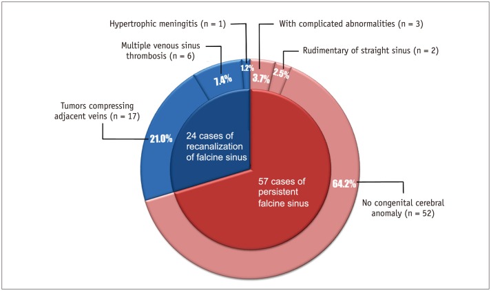
Fig. 1 Associated imaging findings in subjects with falcine sinuses of different clinical types.
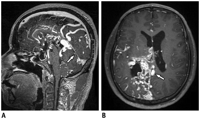
Fig. 2 43-year-old male with multiple cerebral abnormalities.A. Sagittal contrast-enhanced MR image demonstrated absence of splenium of corpus callosum and vascular malformations in corpus callosum and lateral ventricles. Straight vessel was narrow (arrow), with two vessels originating from anterior part and connecting to SSS. B. Axial contrast-enhanced MR image showed that above vessels were in posterior part of falx cerebri (arrow). Vascular malformations were mainly located near right lateral ventricle. MR = magnetic resonance, SSS = superior sagittal sinus
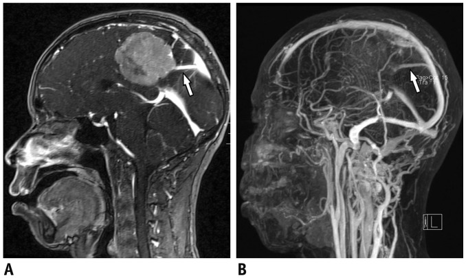
Fig. 3 34-year-old male with meningioma next to falx cerebri.A. Sagittal contrast-enhanced MR image demonstrated close relationship of tumor near middle falx cerebri to falcine sinus (arrow). Straight sinus was normal. B. MR venography image showed arch-like vessel connecting anterior part of straight sinus and posterior part of SSS (arrow).
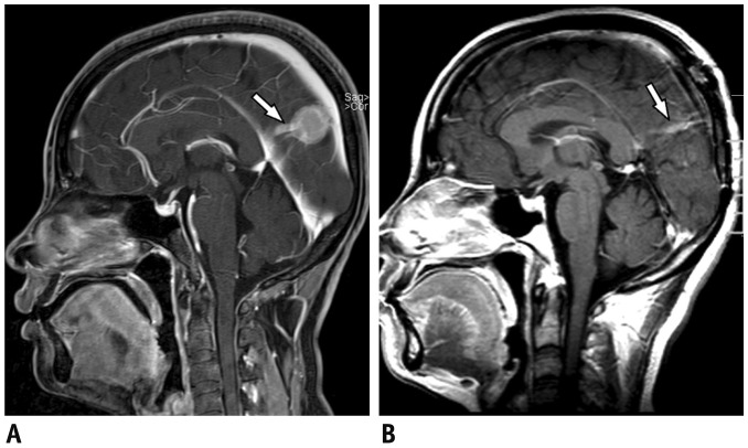
Fig. 4 35-year-old female with meningioma next to falx cerebri.A. Sagittal contrast-enhanced MR image showed that small tumor near posterior part of falx cerebri had close relationship with falcine sinus (arrow). Corpus callosum and straight sinus appeared normal. B. Sagittal contrast-enhanced MR image showed parietal bone defect after removal of tumor; stick-like falcine sinus was preserved (arrow).
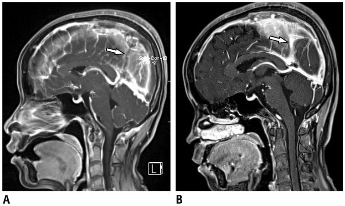
Fig. 5 32-year-old female with multiple venous sinus thrombi.A. Sagittal contrast-enhanced MR images demonstrated defuse thrombosis in SSS, straight sinus and torcular, herophili. Falcine sinus was filled with thrombus (arrow) with distinguishable edge. B. Sagittal contrast-enhanced MR image showed partial absorption of thrombi in SSS and torcular herophili after operation. Thrombi in falcine sinus and straight sinus disappeared and falcine sinus was identified (arrow).
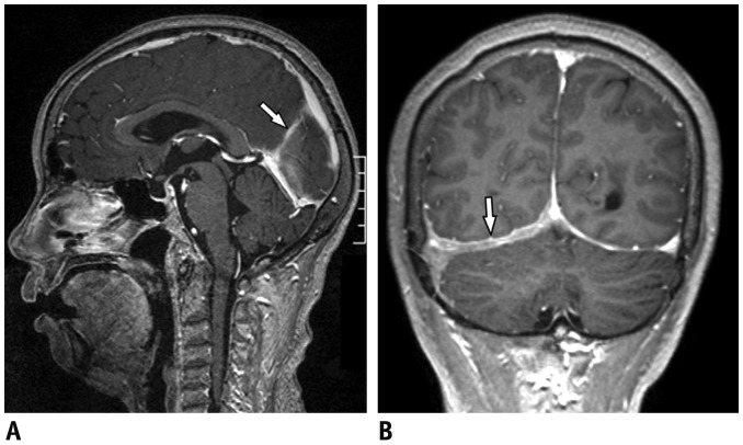
Fig. 6 26-year-old female with hypertrophic meningitis.A. Sagittal contrast-enhanced MR image demonstrated stick-like falcine sinus (arrow). B. Coronal contrast-enhanced MR image detected hypertrophic right tentorium of cerebellum (arrow). Right transversal sinus and sigmoid sinus were compressed.
Reference
-
1. Kesava PP. Recanalization of the falcine sinus after venous sinus thrombosis. AJNR Am J Neuroradiol. 1996; 17:1646–1648. PMID: 8896615.2. Desprechins B, Debaere C, Machiels F, Bougatef A, Osteaux M. A vein of Galen aneurysm with an abnormal drain system: MRI findings. Pediatr Radiol. 1995; 25:442–443. PMID: 7491195.
Article3. Ratliff J, Voorhies RM. Arteriovenous fistula with associated aneurysms coexisting with dural arteriovenous malformation of the anterior inferior falx. Case report and review of the literature. J Neurosurg. 1999; 91:303–307. PMID: 10433319.4. Manoj KS, Krishnamoorthy T, Thomas B, Kapilamoorthy TR. An incidental persistent falcine sinus with dominant straight sinus and hypoplastic distal superior sagittal sinus. Pediatr Radiol. 2006; 36:65–67. PMID: 16283283.
Article5. Abe T, Matsumoto K, Kiyota K, Tanaka H. Vein of Galen aneurysmal malformation in an adult: a case report. Surg Neurol. 1996; 45:39–43. PMID: 9190697.
Article6. Bartels RH, Merx JL, van Overbeeke JJ. Falcine sinus and occipital encephalocele: a magnetic resonance venography study. J Neurosurg. 1998; 89:738–741. PMID: 9817410.
Article7. Kim MS, Lee GJ. Two cases with persistent falcine sinus as congenital variation. J Korean Neurosurg Soc. 2010; 48:82–84. PMID: 20717519.
Article8. Reddy AT, Hedlund GL, Percy AK. Enlarged parietal foramina: association with cerebral venous and cortical anomalies. Neurology. 2000; 54:1175–1178. PMID: 10720293.
Article9. Sener RN. Association of persistent falcine sinus with different clinicoradiologic conditions: MR imaging and MR angiography. Comput Med Imaging Graph. 2000; 24:343–348. PMID: 11008182.
Article10. Cai CQ, Zhang QJ, Yang WD, Wang CX, Shen CH. Neuroimages of persistent falcine sinus in children. World J Pediatr. 2009; 5:63–64. PMID: 19172336.
Article11. Yang CA, Peng SS, Hsieh WS, Tsao PN, Chen CY, Chou HC. Large parietal midline defect with unusual ridge-like structure at the rim and persistent falcine sinus. Childs Nerv Syst. 2013; 29:1069–1072. PMID: 23559396.
Article12. Strub WM, Leach JL, Tomsick TA. Persistent falcine sinus in an adult: demonstration by MR venography. AJNR Am J Neuroradiol. 2005; 26:750–751. PMID: 15814916.13. Kashimura H, Arai H, Ogasawara K, Ogawa A. Persistent falcine sinus associated with obstruction of the superior sagittal sinus caused by meningioma--case report. Neurol Med Chir (Tokyo). 2007; 47:83–84. PMID: 17317947.
Article14. Smith A, Choudhary AK. Prevalence of persistent falcine sinus as an incidental finding in the pediatric population. AJR Am J Roentgenol. 2014; 203:424–425. PMID: 25055280.
Article15. Ryu CW. Persistent falcine sinus: is it really rare? AJNR Am J Neuroradiol. 2010; 31:367–369. PMID: 19779000.
Article
- Full Text Links
-
- Actions
-
Cited
- CITED
-
- Close
- Share
-
- Similar articles
-
- Dynamic Contrast-Enhanced MR Imaging of Tietze’s Syndrome: a Case Report
- Characteristics of Magnetic Resonance(M.R.) and Comprehension of its Imaging Mechanism
- Recent development of diagnostic imaging of hepatocellular carcinoma
- Molecular MR Imaging
- MR Images of Spontaneously Involuted Atretic Cephalocele Concomitant with Persistent Falcine Sinus in an Adult
