Recent development of diagnostic imaging of hepatocellular carcinoma
- Affiliations
-
- 1Department of Radiology and Research Institute of Radiological Science, Yonsei University Severance Hospital, Seoul, Korea. kimnex@yuhs.ac.kr
- KMID: 2193466
- DOI: http://doi.org/10.5124/jkma.2013.56.11.948
Abstract
- Various imaging examinations, such as ultrasonography (US), computed tomography (CT), and magnetic resonance imaging (MRI) have played important roles in the evaluation of hepatocellular carcinoma (HCC). Recent advances in US include contrast-enhanced examinations, fusion imaging, and elastography. For CT, various radiation reduction techniques as well as perfusion imaging and dual-energy examination techniques have been developed. For MRI, hepatocyte-specific contrast-enhanced imaging improved the accuracy of examinations by detection of smaller tumors and better characterization of equivocal lesions. Recent MRI developments also include diffusion-weighted imaging, perfusion imaging, and MR elastography. Newer imaging techniques will increase the roles of imaging examinations in the evaluation of HCC.
Keyword
MeSH Terms
Figure
-
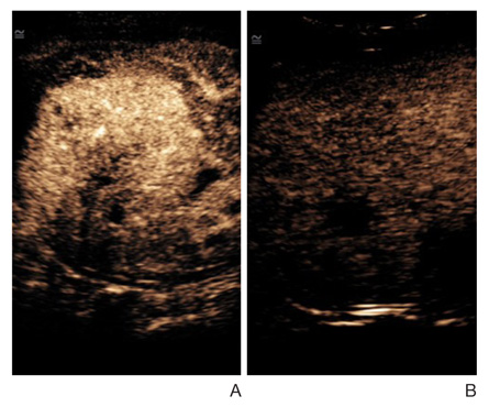
Figure 1 Contrast-enhanced sonography of hepatocellular carcinoma. (A) Arterial phase image obtained 16 seconds after the injection of contrast material shows increased enhancement of the tumor. (B) Delayed phase image obtained at 2 minutes 21 seconds after the injection of the contrast material showed decreased enhancement (washout) of the tumor than surrounding the parenchyma.
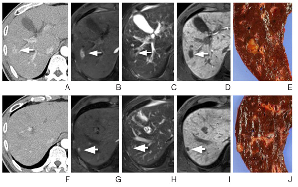
Figure 2 Detection of an additional hepatocellular carcinoma by magnetic resonance imaging (MRI) in a preoperative examination of 60-year-old man. (A-D) Preoperative computed tomography (CT) and MRI show a small hepatocellular carcinoma (arrows). (F-J) At a different level, no lesion was seen on a CT exam (F). However, a small hepatocellular carcinoma is well visible. The both lesions were confirmed on surgery (E,J).

Figure 3 Fusion imaging of magnetic resonance imaging (MRI) and positron emission tomography (PET)-computed tomography. (A) T1-weighted MRI shows an infiltrative tumor (long white arrow) with portal venous thrombosis (short black arrow). (B) Portal venous phase image shows washout of contrast enhancement indicating a tumor thrombosis (arrows) filled in the right portal trunk. (C) T2-weighted images show hyperintense tumor thrombosis and the infiltrative tumor (arrows). (D) Fusion image of transverse T1-weighted magnetic resonance and fluorodeoxyglucose (FDG)-PET exams show increased uptake in the tumor (long white arrow) and in the tumor thrombosis (short black arrow). (E) Fusion image of coronal T2-weighted MRI and FDG-PET exams show increased uptake in the primary tumor (arrow) and absence of extrahepatic metastasis.
Reference
-
1. Singal A, Volk ML, Waljee A, Salgia R, Higgins P, Rogers MA, Marrero JA. Meta-analysis: surveillance with ultrasound for early-stage hepatocellular carcinoma in patients with cirrhosis. Aliment Pharmacol Ther. 2009; 30:37–47.
Article2. Hennedige T, Venkatesh SK. Imaging of hepatocellular carcinoma: diagnosis, staging and treatment monitoring. Cancer Imaging. 2013; 12:530–547.
Article3. Kim TK, Lee KH, Khalili K, Jang HJ. Hepatocellular nodules in liver cirrhosis: contrast-enhanced ultrasound. Abdom Imaging. 2011; 36:244–263.
Article4. Kudo M. Diagnostic imaging of hepatocellular carcinoma: recent progress. Oncology. 2011; 81:Suppl 1. 73–85.
Article5. Makino Y, Imai Y, Igura T, Ohama H, Kogita S, Sawai Y, Fukuda K, Ohashi H, Murakami T. Usefulness of the multimodality fusion imaging for the diagnosis and treatment of hepatocellular carcinoma. Dig Dis. 2012; 30:580–587.
Article6. Friedrich-Rust M, Ong MF, Herrmann E, Dries V, Samaras P, Zeuzem S, Sarrazin C. Real-time elastography for noninvasive assessment of liver fibrosis in chronic viral hepatitis. AJR Am J Roentgenol. 2007; 188:758–764.
Article7. Gheorghe L, Iacob S, Iacob R, Dumbrava M, Becheanu G, Herlea V, Gheorghe C, Lupescu I, Popescu I. Real time elastography: a non-invasive diagnostic method of small hepatocellular carcinoma in cirrhosis. J Gastrointestin Liver Dis. 2009; 18:439–446.8. D'Onofrio M, Gallotti A, Mucelli RP. Tissue quantification with acoustic radiation force impulse imaging: measurement repeatability and normal values in the healthy liver. AJR Am J Roentgenol. 2010; 195:132–136.9. Kwon HJ, Kang MJ, Cho JH, Oh JY, Nam KJ, Han SY, Lee SW. Acoustic radiation force impulse elastography for hepatocellular carcinoma-associated radiofrequency ablation. World J Gastroenterol. 2011; 17:1874–1878.
Article10. Davarpanah AH, Weinreb JC. The role of imaging in hepatocellular carcinoma: the present and future. J Clin Gastroenterol. 2013; 47:Suppl. S7–S10.11. Heye T, Nelson RC, Ho LM, Marin D, Boll DT. Dual-energy CT applications in the abdomen. AJR Am J Roentgenol. 2012; 199:5 Suppl. S64–S70.
Article12. Goh V, Ng QS, Miles K. Computed tomography perfusion imaging for therapeutic assessment: has it come of age as a biomarker in oncology? Invest Radiol. 2012; 47:2–4.13. Golfieri R, Renzulli M, Lucidi V, Corcioni B, Trevisani F, Bolondi L. Contribution of the hepatobiliary phase of Gd-EOB-DTPA-enhanced MRI to Dynamic MRI in the detection of hypovascular small (≤ 2 cm) HCC in cirrhosis. Eur Radiol. 2011; 21:1233–1242.
Article14. Cha DI, Lee MW, Kim YK, Kim SH, Park HJ, Rhim H, Lim HK. Assessing patients with hepatocellular carcinoma meeting the milan criteria: is liver 3 tesla MR with gadoxetic acid necessary in addition to liver CT? J Magn Reson Imaging. 2013; 09. 30. [Epub]. DOI: 10.1002/jmri.24237.
Article15. Bharwani N, Koh DM. Diffusion-weighted imaging of the liver: an update. Cancer Imaging. 2013; 13:171–185.
Article16. Nasu K, Kuroki Y, Tsukamoto T, Nakajima H, Mori K, Minami M. Diffusion-weighted imaging of surgically resected hepatocellular carcinoma: imaging characteristics and relationship among signal intensity, apparent diffusion coefficient, and histopathologic grade. AJR Am J Roentgenol. 2009; 193:438–444.
Article17. Suh YJ, Kim MJ, Choi JY, Park MS, Kim KW. Preoperative prediction of the microvascular invasion of hepatocellular carcinoma with diffusion-weighted imaging. Liver Transpl. 2012; 18:1171–1178.
Article18. Bonekamp S, Li Z, Geschwind JF, Halappa VG, Corona-Villalobos CP, Reyes D, Pawlik TM, Bonekamp D, Eng J, Kamel IR. Unresectable hepatocellular carcinoma: MR imaging after intraarterial therapy. Part I. Identification and validation of volumetric functional response criteria. Radiology. 2013; 268:420–430.
Article19. Jarnagin WR, Schwartz LH, Gultekin DH, Gonen M, Haviland D, Shia J, D'Angelica M, Fong Y, Dematteo R, Tse A, Blumgart LH, Kemeny N. Regional chemotherapy for unresectable primary liver cancer: results of a phase II clinical trial and assessment of DCE-MRI as a biomarker of survival. Ann Oncol. 2009; 20:1589–1595.
Article20. Venkatesh SK, Yin M, Glockner JF, Takahashi N, Araoz PA, Talwalkar JA, Ehman RL. MR elastography of liver tumors: preliminary results. AJR Am J Roentgenol. 2008; 190:1534–1540.
Article
- Full Text Links
-
- Actions
-
Cited
- CITED
-
- Close
- Share
-
- Similar articles
-
- Imaging Diagnosis of Hepatocellular Carcinoma
- Diagnosis of hepatocellular carcinoma: Which MRI contrast agent? Which diagnostic criteria?
- Hepatocarcinogenesis in liver cirrhosis: imaging diagnosis
- Atypical Growth & Development of Classic Hepatocellular Carcinoma
- Intraductal malignant tumors in the liver mimicking cholangiocarcinoma: Imaging features for differential diagnosis
