Korean J Radiol.
2018 Jun;19(3):417-424. 10.3348/kjr.2018.19.3.417.
The Diagnostic Reproducibility of Tomosynthesis for the Correlation between Acromiohumeral Distance and Rotator Cuff Size or Type
- Affiliations
-
- 1Department of Radiology, Hanyang University Hospital, Seoul 04763, Korea. radsh@hanyang.ac.kr
- 2Department of Orthopedics, Hanyang University Hospital, Seoul 04763, Korea.
- 3Department of Rheumatology, Hanyang University Hospital, Seoul 04763, Korea.
- KMID: 2410812
- DOI: http://doi.org/10.3348/kjr.2018.19.3.417
Abstract
OBJECTIVE
To correlate the acromiohumeral distance (AHD) using tomosynthesis and rotator cuff (RC) pathology and various anatomical indices and to assess the diagnostic reproducibility of tomosynthesis for the evaluation of subacromial impingement.
MATERIALS AND METHODS
A retrospective review of 63 patients with clinically suspected subacromial impingement was conducted. Two musculoskeletal radiologists independently measured the following quantitative data: the AHD on plain radiographs and the AHD at three compartments (anterior, middle, and posterior) using tomosynthesis, computed tomography (CT) arthrography, or magnetic resonance (MR) arthrography. To investigate the association between the AHD and RC pathology and various anatomical indices, we reviewed the arthroscopic operation record as the referenced standard.
RESULTS
The size of rotator cuff tear (RCT) in full-thickness tears displayed a significant inverse correlation with the middle and the posterior tomosynthetic AHDs (p < 0.05). The results of an ANOVA revealed that the middle tomosynthetic AHD retained a significant association with the type of RCT (p = 0.042), and the posterior tomosynthetic AHD retained significance for the size of RCT in a full-thickness tear (p = 0.024). The inter-modality correlation exhibited significant agreement especially among the plain radiography, tomosynthesis, and CT or MR arthrography (p < 0.05). The intraobserver and interobserver correlation coefficients (ICCs) displayed excellent agreement (ICC = 0.896-0.983). The humeral head diameter and glenoid height were significantly correlated with patient height and weight.
CONCLUSION
Acromiohumeral distance measurement using tomosynthesis is reproducible compared with other modalities.
MeSH Terms
Figure
-
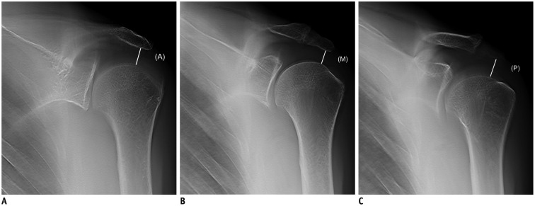
Fig. 1 Schema for measuring AHD on tomosynthesis (A-C).(A); anterior tomosynthetic AHD, (M); middle tomosynthetic AHD, (P); posterior tomosynthetic AHD. AHD = acromiohumeral distance
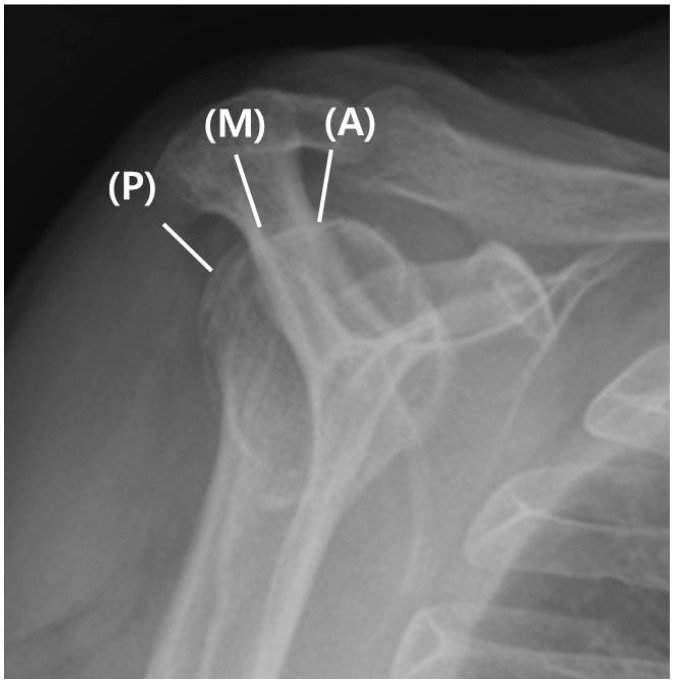
Fig. 2 Schema for measuring AHD on correlated scapular Y view.
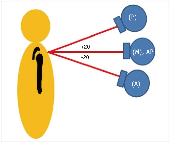
Fig. 3 Schema for X-ray tube position according to AHD measurement on plain radiography and tomosynthesis.AP = anterior-posterior
Reference
-
1. Michener LA, McClure PW, Karduna AR. Anatomical and biomechanical mechanisms of subacromial impingement syndrome. Clin Biomech (Bristol, Avon). 2003; 18:369–379.
Article2. Neer CS 2nd. Anterior acromioplasty for the chronic impingement syndrome in the shoulder: a preliminary report. J Bone Joint Surg Am. 1972; 54:41–50. PMID: 5054450.3. Neer CS 2nd. Impingement lesions. Clin Orthop Relat Res. 1983; 173:70–77.
Article4. Saupe N, Pfirrmann CW, Schmid MR, Jost B, Werner CM, Zanetti M. Association between rotator cuff abnormalities and reduced acromiohumeral distance. AJR Am J Roentgenol. 2006; 187:376–382. PMID: 16861541.
Article5. Leong HT, Tsui S, Ying M, Leung VY, Fu SN. Ultrasound measurements on acromio-humeral distance and supraspinatus tendon thickness: test-retest reliability and correlations with shoulder rotational strengths. J Sci Med Sport. 2012; 15:284–291. PMID: 22209419.
Article6. Ellman H, Hanker G, Bayer M. Repair of the rotator cuff. End-result study of factors influencing reconstruction. J Bone Joint Surg Am. 1986; 68:1136–1144. PMID: 3771595.
Article7. Norwood LA, Barrack R, Jacobson KE. Clinical presentation of complete tears of the rotator cuff. J Bone Joint Surg Am. 1989; 71:499–505. PMID: 2703509.
Article8. Lei J, Yang P, Zhang L, Wang Y, Yang K. Diagnostic accuracy of digital breast tomosynthesis versus digital mammography for benign and malignant lesions in breasts: a meta-analysis. Eur Radiol. 2014; 24:595–602. PMID: 24121712.
Article9. Haas BM, Kalra V, Geisel J, Raghu M, Durand M, Philpotts LE. Comparison of tomosynthesis plus digital mammography and digital mammography alone for breast cancer screening. Radiology. 2013; 269:694–700. PMID: 23901124.
Article10. Vikgren J, Zachrisson S, Svalkvist A, Johnsson AA, Boijsen M, Flinck A, et al. Comparison of chest tomosynthesis and chest radiography for detection of pulmonary nodules: human observer study of clinical cases. Radiology. 2008; 249:1034–1041. PMID: 18849504.
Article11. Göthlin JH, Geijer M. The utility of digital linear tomosynthesis imaging of total hip joint arthroplasty with suspicion of loosening: a prospective study in 40 patients. Biomed Res Int. 2013; 2013:594–631.
Article12. Aoki T, Fujii M, Yamashita Y, Takahashi H, Oki H, Hayashida Y, et al. Tomosynthesis of the wrist and hand in patients with rheumatoid arthritis: comparison with radiography and MRI. AJR Am J Roentgenol. 2014; 202:386–390. PMID: 24450681.
Article13. Ottenin MA, Jacquot A, Grospretre O, Noël A, Lecocq S, Louis M, et al. Evaluation of the diagnostic performance of tomosynthesis in fractures of the wrist. AJR Am J Roentgenol. 2012; 198:180–186. PMID: 22194495.
Article14. Hayashi D, Xu L, Roemer FW, Hunter DJ, Li L, Katur AM, et al. Detection of osteophytes and subchondral cysts in the knee with use of tomosynthesis. Radiology. 2012; 263:206–215. PMID: 22438445.
Article15. Geijer M, Börjesson AM, Göthlin JH. Clinical utility of tomosynthesis in suspected scaphoid fracture. A pilot study. Skeletal Radiol. 2011; 40:863–867. PMID: 21057785.
Article16. Harris G, Bou–Haidar P, Harris C. Adhesive capsulitis: review of imaging and treatment. J Med Imaging Radiat Oncol. 2013; 57:633–643. PMID: 24283550.
Article17. Walton J, Mahajan S, Paxinos A, Marshall J, Bryant C, Shnier R, et al. Diagnostic values of tests for acromioclavicular joint pain. J Bone Joint Surg Am. 2004; 86-A:807–812. PMID: 15069148.
Article18. Pearsall AW 4th, Bonsell S, Heitman RJ, Helms CA, Osbahr D, Speer KP. Radiographic findings associated with symptomatic rotator cuff tears. J Shoulder Elbow Surg. 2003; 12:122–127. PMID: 12700562.
Article19. Pennington RG, Bottomley NJ, Neen D, Brownlow HC. Radiological features of osteoarthritis of the acromiclavicular joint and its association with clinical symptoms. J Orthop Surg (Hong Kong). 2008; 16:300–302. PMID: 19126894.
Article20. Bonsell S, Pearsall AW 4th, Heitman RJ, Helms CA, Major NM, Speer KP. The relationship of age, gender, and degenerative changes observed on radiographs of the shoulder in asymptomatic individuals. J Bone Joint Surg Br. 2000; 82:1135–1139. PMID: 11132273.
Article21. Gruber G, Bernhardt GA, Clar H, Zacherl M, Glehr M, Wurnig C. Measurement of the acromiohumeral interval on standardized anteroposterior radiographs: a prospective study of observer variability. J Shoulder Elbow Surg. 2010; 19:10–13. PMID: 19556147.
Article22. Okkalides D, Fotakis M. Patient effective dose resulting from radiographic examinations. Br J Radiol. 1994; 67:564–572. PMID: 8032810.
Article23. Ha AS, Lee AY, Hippe DS, Chou SH, Chew FS. Digital tomosynthesis to evaluate fracture healing: prospective comparison with radiography and CT. AJR Am J Roentgenol. 2015; 205:136–141. PMID: 26102392.
Article24. Tapiovaara M, Siiskonen T. PCXMC. A Monte Carlo program for calculating patient doses in medical x-ray examinations. STUK-A231. 2nd ed. Helsinki: Radiation and Nuclear Safety Authority;2008.25. Appendix f: pcxmc--a pc-based monte carlo program for calculating patient doses in medical x-ray examinations. JICRU. 2005; 5:100–102.26. Werner CM, Conrad SJ, Meyer DC, Keller A, Hodler J, Gerber C. Intermethod agreement and interobserver correlation of radiologic acromiohumeral distance measurements. J Shoulder Elbow Surg. 2008; 17:237–240. PMID: 18162412.27. Roche C, Angibaud L, Flurin PH, Wright T, Fulkerson E, Zuckerman J. Anatomic validation of an “anatomic” shoulder system. Bull Hosp Jt Dis. 2006; 63:93–97. PMID: 16878825.28. Snyder SJ. Shoulder arthroscopy. 2nd ed. Philadelphia: Lippincott William & Wilkins;2002.29. Ellman H. Diagnosis and treatment of incomplete rotator cuff tears. Clin Orthop Relat Res. 1990; 254:64–74.
Article30. Hamada K, Fukuda H, Mikasa M, Kobayashi Y. Roentgenographic findings in massive rotator cuff tears. A long-term observation. Clin Orthop Relat Res. 1990; 254:92–96.
Article31. Huang LF, Rubin DA, Britton CA. Greater tuberosity changes as revealed by radiography: lack of clinical usefulness in patients with rotator cuff disease. AJR Am J Roentgenol. 1999; 172:1381–1388. PMID: 10227521.
Article32. Petersson CJ, Redlund-Johnell I. The subacromial space in normal shoulder radiographs. Acta Orthop Scand. 1984; 55:57–58. PMID: 6702430.
Article33. Hébert LJ, Moffet H, Dufour M, Moisan C. Acromiohumeral distance in a seated position in persons with impingement syndrome. J Magn Reson Imaging. 2003; 18:72–79. PMID: 12815642.
Article
- Full Text Links
-
- Actions
-
Cited
- CITED
-
- Close
- Share
-
- Similar articles
-
- Factors Influencing the Restoration of Acromiohumeral Distance of Immediate Postoperative Period in Patients Who Have Rotator Cuff Repair Surgery with Large-to-Massive Rotator Cuff Tears
- Clinical Outcome of Arthroscopic Partial Repair of Large to Massive Posterosuperior Rotator Cuff Tears: Medialization of the Attachment Site of the Rotator Cuff Tendon
- Plain Radiographic Findings of a Supraspinatus Tear: Correlation with MR Findings
- Quantitative T2 Mapping of Articular Cartilage of the Glenohumeral Joint at 3.0T in Rotator Cuff Disease Patients: the Evaluation of Degenerative Change of Cartilage
- Primary Repair in Tears Affecting Two or More Rotator Cuff Tendons
