Quantitative T2 Mapping of Articular Cartilage of the Glenohumeral Joint at 3.0T in Rotator Cuff Disease Patients: the Evaluation of Degenerative Change of Cartilage
- Affiliations
-
- 1Department of Radiology, Jeju National University Hospital, Jeju-si, Korea. we1977@naver.com
- KMID: 2459877
- DOI: http://doi.org/10.13104/imri.2019.23.3.228
Abstract
- PURPOSE
The aim of this study is to evaluate the T2 value of the articular cartilage of the glenohumeral joint in rotator cuff disease displayed on 3.0T MRI and to apply it in clinical practice.
MATERIALS AND METHODS
This study involved sixty-two patients who underwent shoulder MRI containing T2 mapping. The mean T2 value was measured by placing a free hand ROI over the glenoid or humeral cartilage from the bone-cartilage interface to the articular surface on three consecutive, oblique coronal images. The drawn ROI was subsequently divided into superior and inferior segments. The assessed mean T2 values of the articular cartilage of the glenohumeral joint were compared and evaluated based on the degree of rotator cuff tear, the degree of fatty atrophy of the rotator cuff, and the acromiohumeral distance.
RESULTS
ICC values between two readers indicated moderate or good reproducibility. The mean T2 value for the articular cartilage of the glenoid and humeral head cartilage failed to show any significant difference based on the degree of rotator cuff tear. However, the mean T2 values of articular cartilage, based on fatty atrophy, tended to be higher in fatty atrophy 3 or fatty atrophy 4 groups while some sub-regions displayed significantly higher mean T2 values. There was no correlation between the acromiohumeral distance and the mean T2 values of the articular cartilage of the glenoid and humeral head.
CONCLUSION
T2 mapping of the glenohumeral joint failed to show any significant difference in quantitative analysis of the degenerative change of the articular cartilage based on the degree of rotator cuff tear. However, it also offers quantitative information on the degenerative change of cartilage of the glenohumeral joint in patients with rotator cuff tear and severe fatty atrophy of the rotator cuff.
Keyword
MeSH Terms
Figure
-

Fig. 1. T2 value measurement (a, b). Three consecutive oblique coronal sections comprising large areas of humeral and glenoid cartilage were selected from the images. (c, d) On color-coded T2 maps comparing the T2-weighted image. ROI was drawn on targeted cartilage of the glenoid and humeral head respectively. The drawn ROI was subsequently divided into superior and inferior segments.
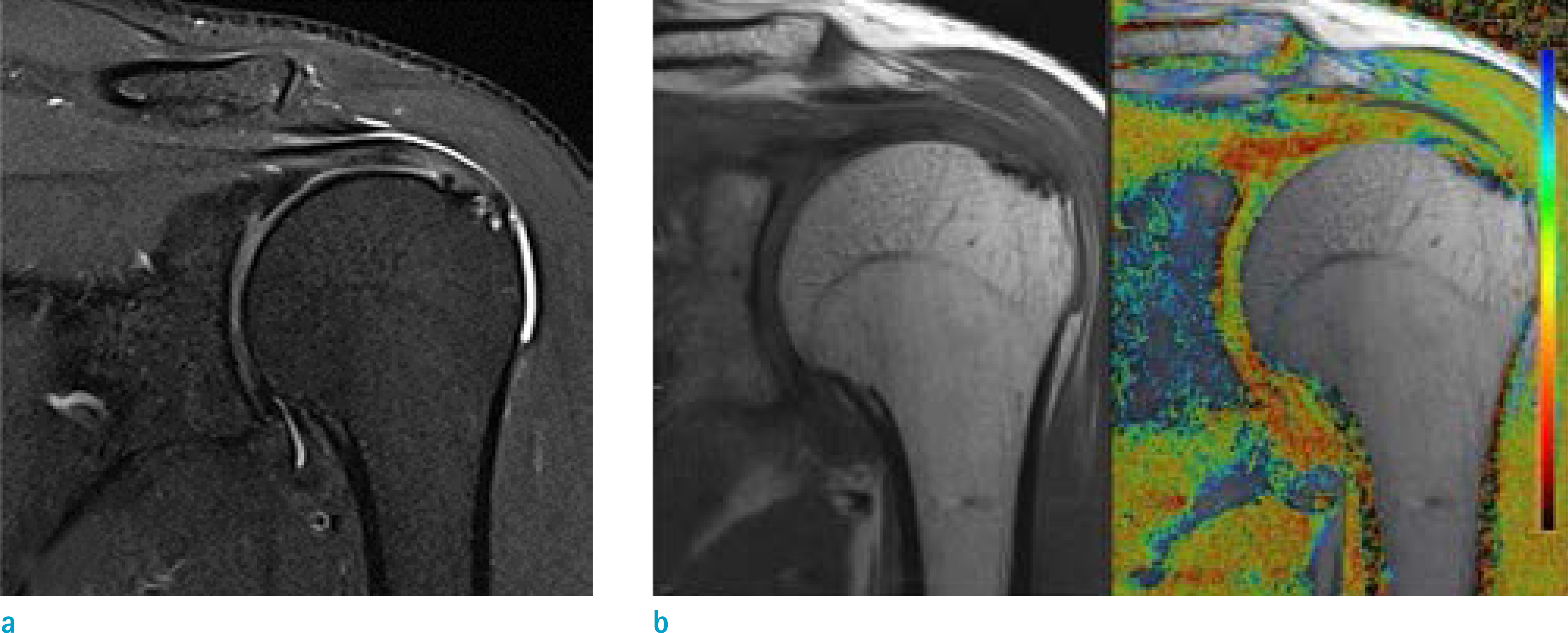
Fig. 2. (a) 46-year-old man with tendinopathy of rotator cuff and Goutallier grade 2. (b) The T2 map of the glenohumeral joint indicates slightly lower T2 value compared to the reported normal range. (The mean T2 value of reader 1; glenoid [41.88 ms], humeral head [41.95 ms], the mean T2 value of reader 2; glenoid [38.60 ms], humeral head [43.30 ms].)
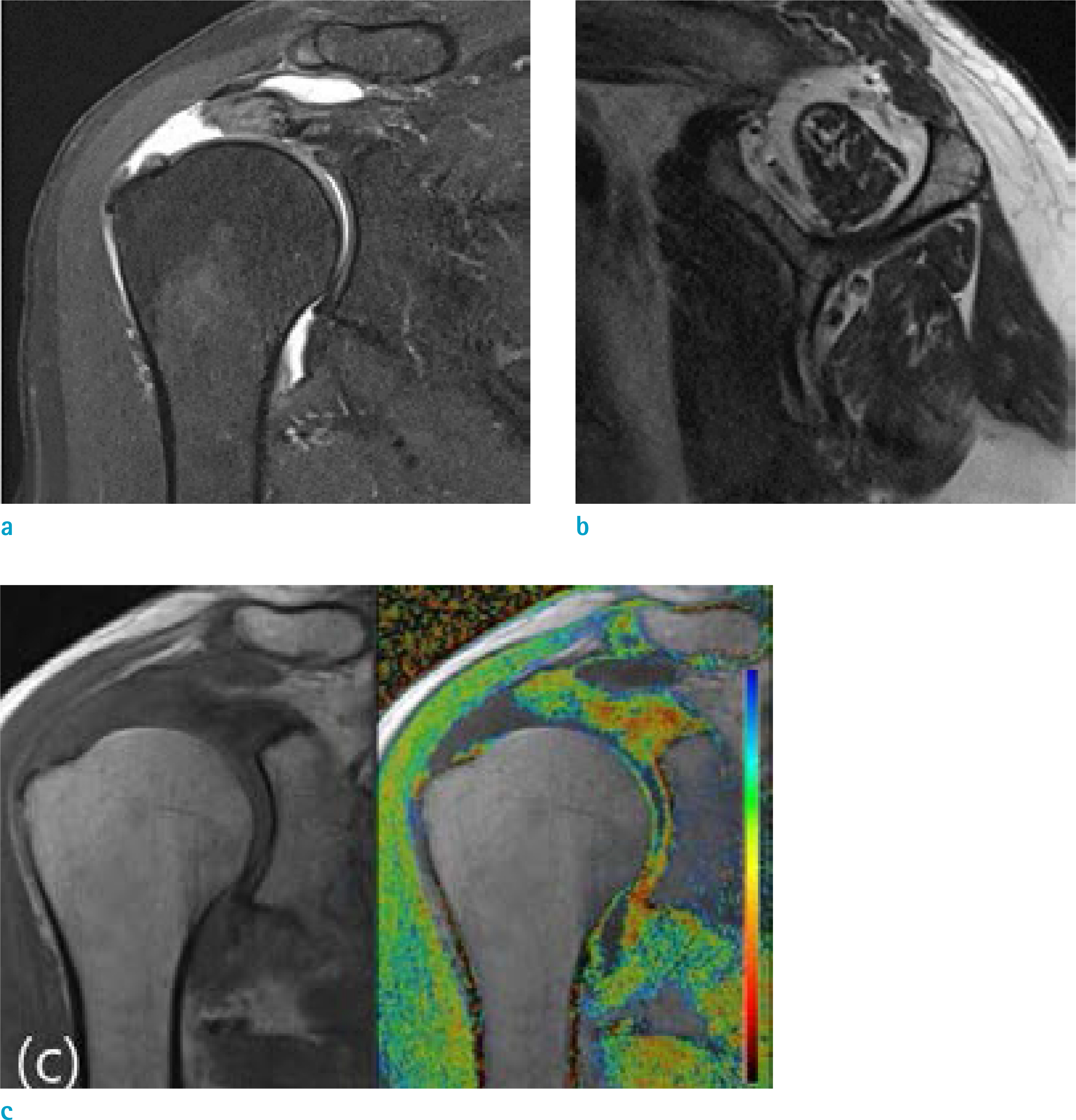
Fig. 3. (a, b) 76-year-old woman with full thickness tear of rotator cuff and Goutallier grade 3. (c) The T2 map of the glenohumeral joint showing higher T2 value compared to the reported normal range. The mean T2 value of glenoid according to reader 2 was similar with the reported normal range. (The mean T2 value of reader 1; glenoid [99.15 ms], humeral head [59.43 ms], the mean T2 value of reader 2; glenoid [47.57 ms], humeral head [56.40 ms])
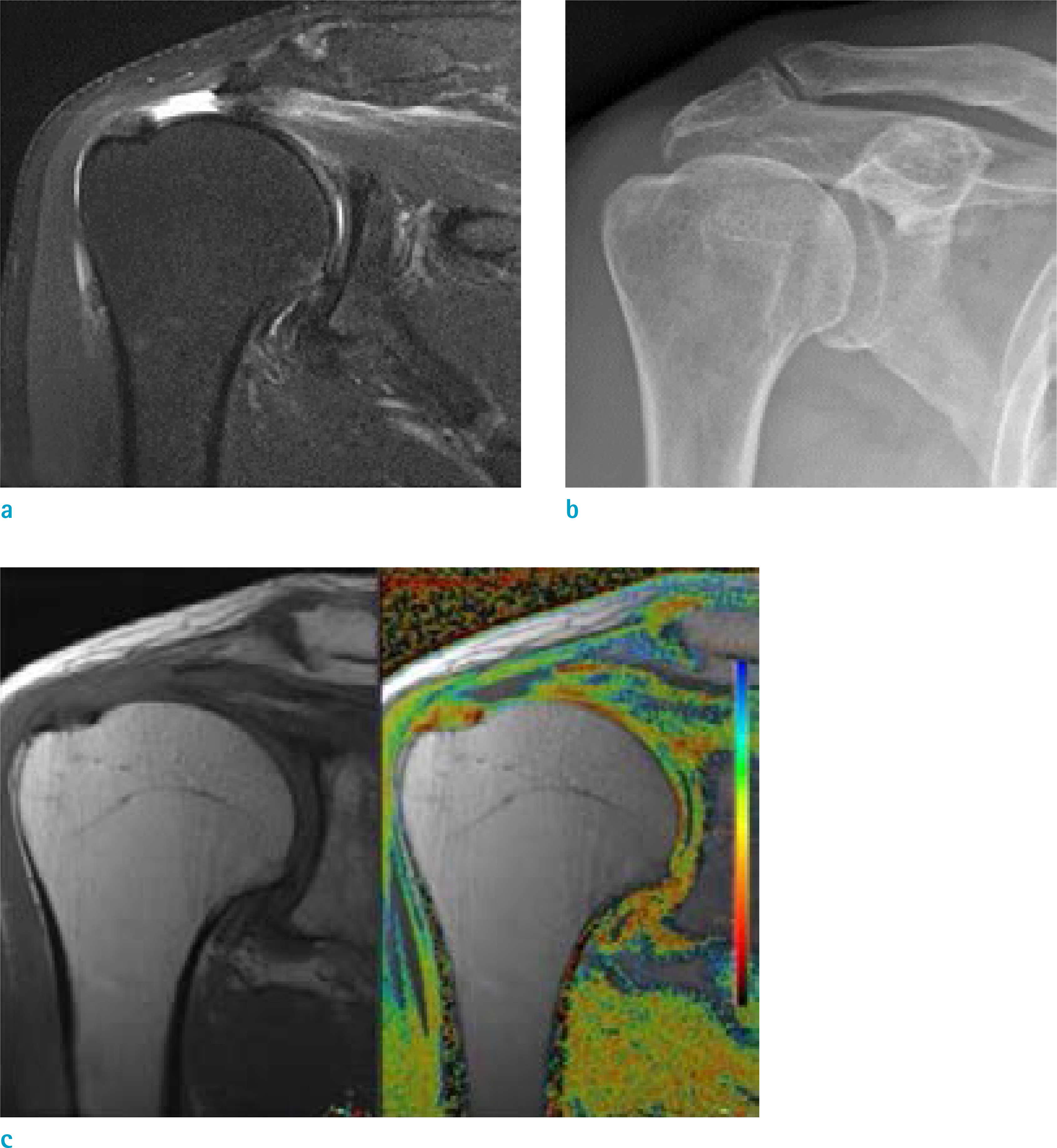
Fig. 4. (a, b) 72-year-old man with full thickness tear of rotator cuff and decreased acromiohumeral distance (AHD; 6.90 cm). (c) The T2 map of the glenohumeral joint indicates similar or slightly lower T2 value compared to the reported normal range. (The mean T2 value of reader 1; glenoid [45.35 ms], humeral head [48.91 ms], the mean T2 value of reader 2; glenoid [47.70 ms], humeral head [41.90 ms])
Reference
-
References
1. Sambandam SN, Khanna V, Gul A, Mounasamy V. Rotator cuff tears: an evidence based approach. World J Orthop. 2015; 6:902–918.
Article2. Mitchell JJ, Warner BT, Horan MP, et al. Comprehensive arthroscopic management of glenohumeral osteoarthritis: preoperative factors predictive of treatment failure. Am J Sports Med. 2017; 45:794–802.
Article3. Jeong HY, Jeon YS, Lee DK, Rhee YG. Rotator cuff tear with early osteoarthritis: how does it affect clinical outcome after large to massive rotator cuff repair? J Shoulder Elbow Surg. 2019; 28:237–243.
Article4. McCauley TR, Recht MP, Disler DG. Clinical imaging of articular cartilage in the knee. Semin Musculoskelet Radiol. 2001; 5:293–304.
Article5. Mosher TJ, Dardzinski BJ. Cartilage MRI T2 relaxation time mapping: overview and applications. Semin Musculoskelet Radiol. 2004; 8:355–368.
Article6. Apprich S, Mamisch TC, Welsch GH, et al. Quantitative T2 mapping of the patella at 3.0T is sensitive to early cartilage degeneration, but also to loading of the knee. Eur J Radiol. 2012; 81:e438–443.
Article7. Dardzinski BJ, Mosher TJ, Li S, Van Slyke MA, Smith MB. Spatial variation of T2 in human articular cartilage. Radiology. 1997; 205:546–550.
Article8. Dunn TC, Lu Y, Jin H, Ries MD, Majumdar S. T2 relaxation time of cartilage at MR imaging: comparison with severity of knee osteoarthritis. Radiology. 2004; 232:592–598.
Article9. Mamisch TC, Trattnig S, Quirbach S, Marlovits S, White LM, Welsch GH. Quantitative T2 mapping of knee cartilage: differentiation of healthy control cartilage and cartilage repair tissue in the knee with unloading–initial results. Radiology. 2010; 254:818–826.
Article10. Koo TK, Li MY. A guideline of selecting and reporting intraclass correlation coefficients for reliability research. J Chiropr Med. 2016; 15:155–163.
Article11. Neer CS 2nd. Replacement arthroplasty for glenohumeral osteoarthritis. J Bone Joint Surg Am. 1974; 56:1–13.
Article12. Chalmers PN, Salazar DH, Steger-May K, et al. Radiographic progression of arthritic changes in shoulders with degenerative rotator cuff tears. J Shoulder Elbow Surg. 2016; 25:1749–1755.
Article13. Ellman H, Harris E, Kay SP. Early degenerative joint disease simulating impingement syndrome: arthroscopic findings. Arthroscopy. 1992; 8:482–487.
Article14. Yamaguchi K, Ditsios K, Middleton WD, Hildebolt CF, Galatz LM, Teefey SA. The demographic and morphological features of rotator cuff disease. A comparison of asymptomatic and symptomatic shoulders. J Bone Joint Surg Am. 2006; 88:1699–1704.15. Maizlin ZV, Clement JJ, Patola WB, et al. T2 mapping of articular cartilage of glenohumeral joint with routine MRI correlation–initial experience. HSS J. 2009; 5:61–66.
Article16. Feeney MS, O'Dowd J, Kay EW, Colville J. Glenohumeral articular cartilage changes in rotator cuff disease. J Shoulder Elbow Surg. 2003; 12:20–23.
Article17. Fitzgerald M, Lawler SM, Lowe JT, Nelson R, Mantell MT, Jawa A. Computed tomography underestimates rotator cuff pathology in patients with glenohumeral osteoarthritis. J Shoulder Elbow Surg. 2018; 27:1451–1455.
Article18. Tran G, Cowling P, Smith T, et al. What imaging-detected pathologies are associated with shoulder symptoms and their persistence? A systematic literature review. Arthritis Care Res (Hoboken). 2018; 70:1169–1184.
Article19. Eagle S, Potter HG, Koff MF. Morphologic and quantitative magnetic resonance imaging of knee articular cartilage for the assessment of posttraumatic osteoarthritis. J Orthop Res. 2017; 35:412–423.
Article20. Link TM, Stahl R, Woertler K. Cartilage imaging: motivation, techniques, current and future significance. Eur Radiol. 2007; 17:1135–1146.
Article21. Kijowski R, Blankenbaker DG, Munoz Del Rio A, Baer GS, Graf BK. Evaluation of the articular cartilage of the knee joint: value of adding a T2 mapping sequence to a routine MR imaging protocol. Radiology. 2013; 267:503–513.
Article22. Kester BS, Carpenter PM, Yu HJ, et al. T1rho/T2 mapping and histopathology of degenerative cartilage in advanced knee osteoarthritis. World J Orthop. 2017; 8:350–356.23. Lockard CA, Wilson KJ, Ho CP, Shin RC, Katthagen JC, Millett PJ. Quantitative mapping of glenohumeral cartilage in asymptomatic subjects using 3 T magnetic resonance imaging. Skeletal Radiol. 2018; 47:671–682.24. Kang Y, Choi JA. T2 mapping of articular cartilage of the glenohumeral joint at 3.0 T in healthy volunteers: a feasibility study. Skeletal Radiol. 2016; 45:915–920.25. Lee SY, Park HJ, Kwon HJ, et al. T2 relaxation times of the glenohumeral joint at 3.0 T MRI in patients with and without primary and secondary osteoarthritis. Acta Radiol. 2015; 56:1388–1395.26. Zingman A, Li H, Sundem L, et al. Shoulder arthritis secondary to rotator cuff tear: a reproducible murine model and histopathologic scoring system. J Orthop Res. 2017; 35:506–514.
Article27. Flurin PH, Hardy P, Valenti P, et al. Osteoarthritis after rotator cuff repair: a 10-year follow-up study. Orthop Traumatol Surg Res. 2017; 103:477–481.
Article28. Werner CM, Conrad SJ, Meyer DC, Keller A, Hodler J, Gerber C. Intermethod agreement and interobserver correlation of radiologic acromiohumeral distance measurements. J Shoulder Elbow Surg. 2008; 17:237–240.29. Tashjian RZ. Epidemiology, natural history, and indications for treatment of rotator cuff tears. Clin Sports Med. 2012; 31:589–604.30. Zingg PO, Jost B, Sukthankar A, Buhler M, Pfirrmann CW, Gerber C. Clinical and structural outcomes of nonoperative management of massive rotator cuff tears. J Bone Joint Surg Am. 2007; 89:1928–1934.
Article31. Collin P, Thomazeau H, Walch G, et al. Clinical and structural outcome twenty years after repair of isolated supraspinatus tendon tears. J Shoulder Elbow Surg. 2019; 28:196–202.
Article32. Kim JY, Park JS, Rhee YG. Can preoperative magnetic resonance imaging predict the reparability of massive rotator cuff tears? Am J Sports Med. 2017; 45:1654–1663.
Article33. Kuzel BR, Grindel S, Papandrea R, Ziegler D. Fatty infiltration and rotator cuff atrophy. J Am Acad Orthop Surg. 2013; 21:613–623.
Article34. Berhouet J, Collin P, Benkalfate T, et al. Massive rotator cuff tears in patients younger than 65 years. Epidemiology and characteristics. Orthop Traumatol Surg Res. 2009; 95:S13–18.35. Naimark M, Berliner J, Zhang AL, Davies M, Ma CB, Feeley BT. Prevalence of rotator cuff atrophy and fatty infiltration in patients undergoing total shoulder arthroplasty. J Shoulder Elbow Arthroplasty. 2017; 1:247154921770832–7.
Article36. Iannotti JP, Norris TR. Influence of preoperative factors on outcome of shoulder arthroplasty for glenohumeral osteoarthritis. J Bone Joint Surg Am. 2003; 85:251–258.
Article37. Goutallier D, Le Guilloux P, Postel JM, Radier C, Bernageau J, Zilber S. Acromio humeral distance less than six millimeter: its meaning in full-thickness rotator cuff tear. Orthop Traumatol Surg Res. 2011; 97:246–251.
Article38. Saupe N, Pfirrmann CW, Schmid MR, Jost B, Werner CM, Zanetti M. Association between rotator cuff abnormalities and reduced acromiohumeral distance. AJR Am J Roentgenol. 2006; 187:376–382.
Article39. Nove-Josserand L, Levigne C, Noel E, Walch G. The acromiohumeral interval. A study of the factors influencing its height. Rev Chir Orthop Reparatrice Appar Mot. 1996; 82:379–385.40. Lammentausta E, Multanen J, Nieminen MT. Differences in T2 values of knee cartilage measured with different scanners. Proc Intl Soc Mag Reson Med. 2009; 17:3986.41. Koff MF, Parratte S, Amrami KK, Kaufman KR. Examiner repeatability of patellar cartilage T2 values. Magn Reson Imaging. 2009; 27:131–136.
Article42. Mosher TJ, Zhang Z, Reddy R, et al. Knee articular cartilage damage in osteoarthritis: analysis of MR image biomarker reproducibility in ACRIN-PA 4001 multicenter trial. Radiology. 2011; 258:832–842.
Article43. Glaser C, Mendlik T, Dinges J, et al. Global and regional reproducibility of T2 relaxation time measurements in human patellar cartilage. Magn Reson Med. 2006; 56:527–534.44. Koff MF, Amrami KK, Felmlee JP, Kaufman KR. Bias of cartilage T2 values related to method of calculation. Magn Reson Imaging. 2008; 26:1236–1243.
Article
- Full Text Links
-
- Actions
-
Cited
- CITED
-
- Close
- Share
-
- Similar articles
-
- The Change of Articular Cartilage Thickness of the Knee Joint Related to Age in Korean
- Autogeous Bone-Articular Cartilage stored within Abdominal Wall
- An Investigation of Articular Cartilage Degeneration Induced by Compression-Immobilization and Condylar resection of Knee Joint in Rabbits
- Occult Interpositional Rotator Cuff - an Extremely Rare Case of Traumatic Rotator Cuff Tear
- Current Update of Cartilage Regeneration Using Stem Cells in Osteoarthritis
