Transesophageal Echocardiographic Findings of Ischemic Stroke without Obvious Cardiac Disease
- Affiliations
-
- 1Department of Internal Medicine and Neurology, Chosun University Hospital, Kwangju, Korea.
- KMID: 2410463
- DOI: http://doi.org/10.4250/jkse.1994.2.1.80
Abstract
- To detect the cardiac source of embolism in patient of ischemic stroke of uncertain etiology, biplane transesophageal echocardiography and contrast echocardiography with hand-agitated saline were performed 27 patients(sixteen men and eleven women) of transient ischemic attack and cerebral infarction without definitive cardiac symptom and sign. Transesophageal echocardiography showed potential sources of embolism in nineteen patients (70.4%) including atrial septal aneurysm(n = 9, three of them had patent foramen ovale), spontaneous contrast echo(n = 3), mitral valve prolapse(n= 1), unknown thickening of the tip of the mitral valve(n = 1) and atherosclerotic plaque in descending aorta(n = 7). Thus transesophageal echocardiography and contrast echocardiography identify potential cardiac source of embolism, and provide the rationale of the thrombolytic and anticoagulant therapy in patients with ischemic stroke without obvious cardiac disease.
MeSH Terms
Figure
-
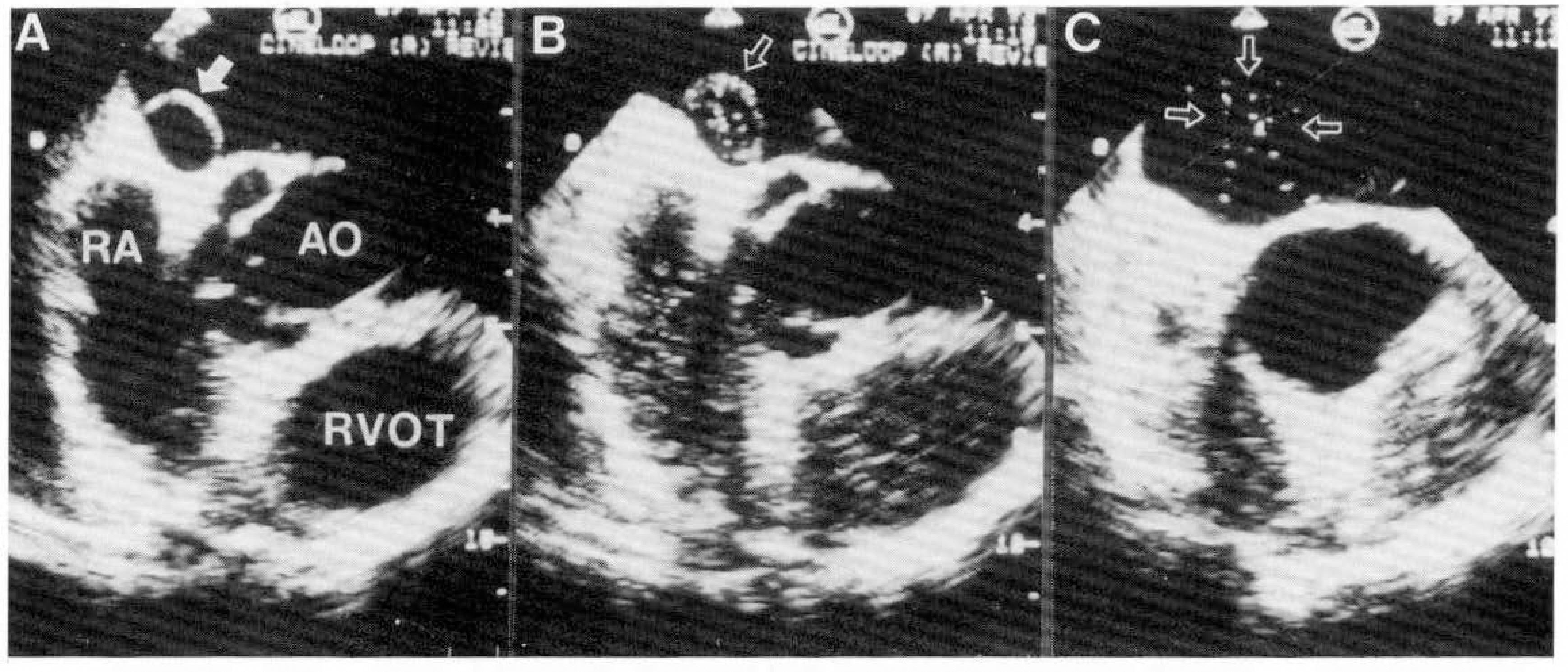
Fig. 1. A; Transesophageal echocardiogram showing an atrial septal aneurysm(Arrow). The atrial septum bulges towards the left atrium. B; After hand-agitated contrast administration contrast fills right atrium (RA), right ventricular outflow tract(RVOT) and atrial septal aneurysm(open arrow). C; The small amount of contrast enters into left atrium(three open arrows), suggesting existing the patent foramen ovale.

Fig. 2. A; M-mode echocardiogram of the right atrium(RA) and interatrial septum showing the smoke-like spontaneous contrast echo in right atrium(RA). B; Longitudinal plane of transesophageal echocardiogram showing spontaneous contrast echo in the left ventricular inflow tract. LA; left atrium, LV; left ventricle.
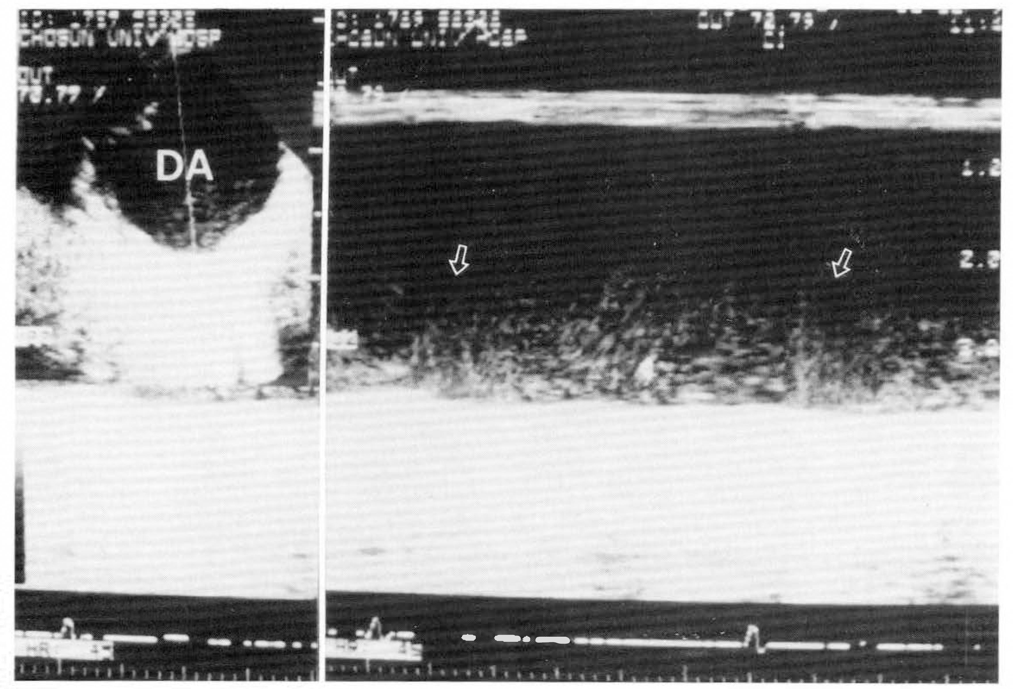
Fig. 3. M-mode echocardiogram of descending aorta(DA) showing spontaneous contrast echo. Note the changes of spontaneous contrast echo during systole(arrows).
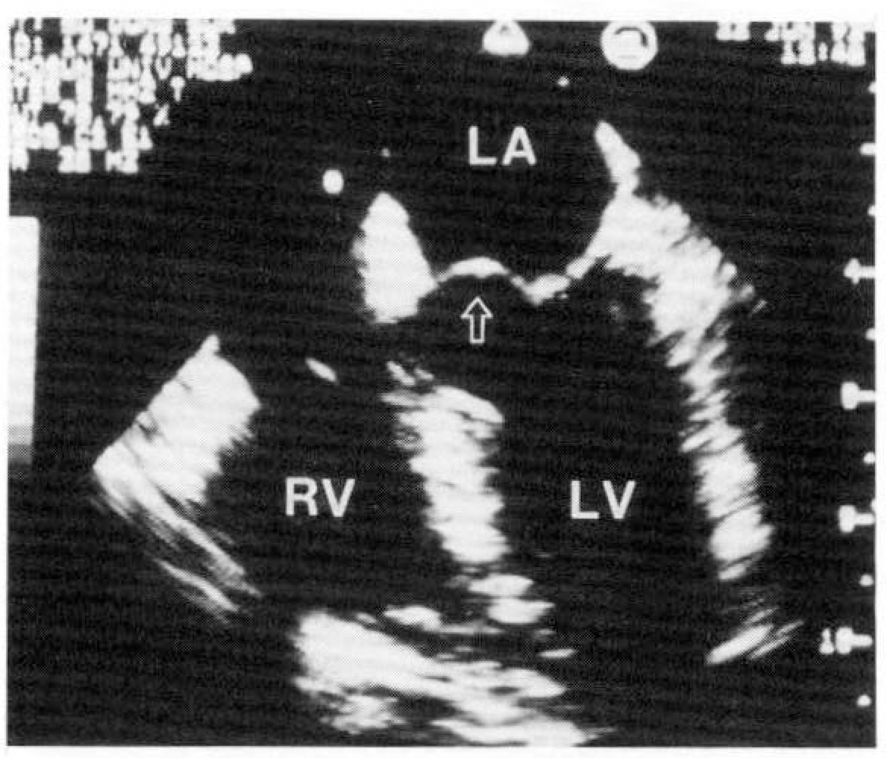
Fig. 4. Transesophageal four-chamber view showing the redundant anterior leaflet protruding into left atrium(LA) during systole(open arrow).

Fig. 5. Transesophageal echocardiogram of mitral valve showing small round thickening of the tip of the both leaflets(arrow). A; diastole, B; systole.

Fig. 6. Transverse plane of transesophageal echocardiogram(A and B) of ascending aorta(ASC. AO) showing the protruding atheroma. Filamentous portion(right side) of atheromatous plaque is moving freely. Longitudinal plane of transesophageal echocardiogram of the same level shows curvilinear atheromatous plaque.

Fig. 7. Transverse plane(A) and longitudinal plane(B) of transesophageal echocardiogram showing a large protruding atherosclerotic plaque in descending aorta.
Reference
-
References
1). Come P, Riley MF, Bivas NK. Role of echocardiography and arrhythmia monitoring in the evaluation of patients with suspected systemic embolism. Ann Neurol. 13:527–531. 1983.2). Nishide M, Irino T, Gotoh M, Naka M, Tsuji K. Cardiac abnormalities in ischemic cerebrovascular disease studied by two-dimensional echocardiography. Stroke. 14:541–545. 1983.
Article3). Good DC, Frank S, Verhulst S, Sharma B. Cardiac abnormalities in stroke patients with negative arteriograms. Stroke. 17:6–11. 1986.
Article4). Randall JL, Thomas B, Tiong-Keat Y, Harlen RG, Dennis C, Ingela S. Enhanced detection of intracardiac sources of cerebral emboli by transesophageal echocardiography. Stroke. 22:734–739. 1991.
Article5). Tei C. Contrast echocardiography. Medicina(Japan). 28:112–119. 1991.6). Seward JB, Bijoy KK, Oh JK, Abel MD, Hughes RW, Edwards WD, Nichols BA, Freeman WK, Tajik AJ. Transesophageal echocardiography: Technique, anatomic correlations, interpretation and clinical applications. Mayo Clinic Proc. 63:649–680. 1986.7). Cerebral Embolism Task Force. Cardiogenic brain embolism: The second report of the cerebral embolism task force. Arch Neurol. 46:727–743. 1989.8). Sherman DG, Dyken ML, Fisher M, Harrison MJG, Hart RG. Antithrombotic therapy for cerebrovascular disorders. Chest. 95(suppl):140S–155S. 1989.
Article9). Hart RG. Cardiogenic embolism to the brain. Lancet. 339:589–594. 1992.
Article10). Biller J, Adams HP Jr, Johnson MR, Kerber RE. Paradoxical cerebral embolism: eight cases. Neurology. 36:1356–1360. 1986.
Article11). Biller J, Johnson MR, Adams HP Jr, Kerber RE, et al. Echocardiographic evaluation of young adults with nonhemorrhagic cerebral infarction. Stroke. 17:608–612. 1986.
Article12). Roberts WC. Aneurysm(redundancy) of the atrial septum (fossa ovale membrane) and prolapse (redundancy) of the mitral valve. Am J Cardiol. 54:1153–1154. 1984.13). Hanley PC, Tajik AJ, Hynes JK, Edwards WD, Reeder GS, Hagler DJ, Seward JB. Diagnosis and classification of atrial septal aneurysm by two-dimensional echocardiography: Report of 80 consecutive cases. Am J Cardiol. 6:1370–1382. 1985.
Article14). Belkin RN, Waugh RA, Kisslo J. Interatrial shunting in atrial septal aneuysm. Am J Cardiol. 57:310–312. 1986.15). Longhini C, Brunazzi C, Musacci G, Caneva M, Bandello A, Bolomini L, Barbiero M, Toselli T, Barbaresi F. Atrial septal aneurysm-echopolycardiographic study. Am J Cardiol. 56:653–666. 1985.16). Belkin RN, Hurwitz BJ, Kisslo J. Atrial septal aneurysm: association with cerebrovascular and peripheral embolic events. Stroke. 18:856–862. 1987.
Article17). Gallet B, Malergue MC, Adams C, Saudemont J, Collot AMC, Druon MC, Hiltgen M. Atrial septal aneurysm-a potential cause of systemic embolism. An echocardiographic study. Br heart J. 53:292–297. 1985.
Article18). Belkin RN, Kisslo J. Atrial septal aneurysm: recognition and clinical relevance. Am heart J. 120:948–957. 1990.
Article19). Schneider B, Hanrath P, Vogel P, Meinertz T. Improved morphologic characterization of atrial septal aneurysm by transesophageal echocardiography: Relation to cerebrovascular events. Am J Cardiol. 16:1000–1009. 1990.
Article20). 장경식·박 일· 국기용·안기완· 홍순표;뇌경 색중 환자에서 난원공 개폰증을 동반한 심방중격류 2혜. 한국심초옴파학회지. 1:131–138. 1993.21). Ramon Castello, Anthony C, Pearson , Labovitz Arthur J. Prevalence and Clinical implications of atrial spontaneous contrast in patients undergoing transesophageal echocardiography. Am J Cardiol. 65:1149–1153. 1990.22). Mikell TL, Asinger RW, Erlsberger J, Anderson RW, Hodges M. Regional stasis of blood in dysfunctional left ventricle: echocardiographic detection and differentiation from early thrombosis. Circulation. 66:755–763. 1982.23). Garcia Fernandez MA, Moreno M, Banuelos F. Two-dimensional echocardiographic identification of blood stasis in the left atrium. Am Heart J. 109:600–601. 1985.24). Iliceto S, Antonelli G, Sorino M, Biasco G, Rizzon P. Dynamic intracavitary left atrial echoes in mitral stenosis. Am J Cardiol. 55:603–606. 1985.
Article25). Erbel R, Stern H, Ehrenthal W, Schreiner G, Treese N, Kramer G, Thelen M, Schweizer P, Meyer J. Detection of spontaneous echocardiographic contrast within the left atrium by transesophageal echocardiography: spontaneous echocardiographic contrast. Clin Cardiol. 9:245–252. 1986.
Article26). Daniel WG, Nellessen V, Schroder E, Nonnast-Daniel B, Bednarski P, Nikutta P, Lichtlen PR. Left atrial spontaneous contrast in mitral valve disease: An indicator for an increased thromboembolic risk. JACC. 11:1204–1211. 1988.27). Kelley RE, Pina I, Lee S-C. Cerebral ischemia and mitral valve prolapse: Case-control study of associated factors. Stroke. 19:443–446. 1988.
Article28). Kouvaras G, Bacoulas G. Association of mitral valve leaflet prolapse with cerebral ischemic events in the young and early middle-aged patient. Q J Med. 55:387–392. 1985.29). Pop G, Sutherland GR, Koudstaal PJ, Sit TW, de Jong G. Transesophageal echocardiography in the detection of intracardiac embolic sources in patients with transient ischemic attacks. Stroke. 21:172–175. 1990.
Article30). Tunick PA, Kronzon I. Protruding atherosclerotic plaque in the aortic arch of patients with systemic embolization: A new finding seen by transesophageal echocardiography. Am Heart J. 120:658–660. 1990.
Article31). Pierre A, Charles D, Christophe T, Dominique H, Marie-Germaine B, Jean-Jacques H. The prevalence of ulcerated plaques in the aortic arch in patients with stroke. N Engl J Med. 326:221–225. 1992.
Article
- Full Text Links
-
- Actions
-
Cited
- CITED
-
- Close
- Share
-
- Similar articles
-
- Transesophageal Echocardiographic Findings in Stroke Subtypes
- Risk Factor Analysis in Patients with Recurrent Cerebral Infarction by Transesophageal Echocardiography
- Transesophageal Echocardiographic Findings Are Independent and Relevant Predictors of Ischemic Stroke in Patients with Nonvalvular Atrial Fibrillation
- Prevalence of the patent foramen ovale in young patients with ischemic cerebrovascular disease: Transesophageal contrast echocardiographic study
- Transesophageal Echocardiographic Evaluation for Potential Sources of Embolism in the Patients with Ischemic Stroke
