J Korean Soc Echocardiogr.
1996 Jul;4(1):29-33. 10.4250/jkse.1996.4.1.29.
Diagnosis of Latent Hypertrophic Obstructive Cardiomyopathy with Dobutamine Stress Echocardiography
- Affiliations
-
- 1Division of Cardiology, Department of Internal Medicine, School of Medicine, Kyung Hee University, Seoul, Korea.
- KMID: 2410403
- DOI: http://doi.org/10.4250/jkse.1996.4.1.29
Abstract
- BACKGROUND
In latent type of hypertrophic obstructive cardiomyopathy, there is no pressure gradient at rest in left ventricular outflow tract(LVOT), but it develops with provocation. Dobutamine increase myocardial contractility and may inducce outflow tract obstruction. To evaluate the usefulness of dobutamine induced outflow tract obstruction as a provocation test, nine patients with latent obstructive cardiomyopathy were studied. METHOD: 680 cases of dobutamine stress echocardiography were reviewed. Nine patients developed late peaking outflow velocity pattern in response to dobutamine infusion(inducible group). Ten patients developed early peaking velocity pattern were included as control group. Left ventricular dimension, outflow tract diameter were measured, and pattern of septal hypertrophy was classified. Changes of peak velocity and acceleration time/ejection time ratio (AT/ET) were measured at rest and peak dose dobutamine.
RESULTS
The peak outflow velocity at rest was not different in both groups(1.49±0.45, 1.18±0.11m/sec). Peak velocity and AT/ET ratio were significantly increased in inducible group(4.2±0.9m/sec, 0.66±0.17), but no significant changes were noted in control group. Patients with inducible group had greater septal thickness, smaller outflow tract diameter and greater prevalence of septal bulge morphology.
CONCLUSION
These results suggest that dobutamine stress Doppler echocardiography could be a useful provocation test to diagnosis of latent obstructive cardiogyopathy.
MeSH Terms
Figure
-
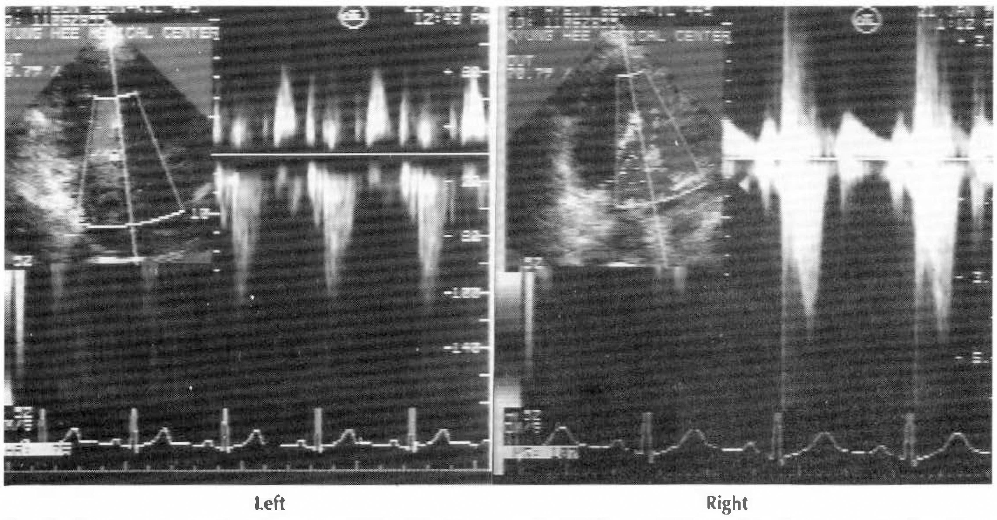
Fig. 1. Continious wave Doppler echocardiographic signals from LVOT at rest(left) and dobutamine infusion(right) in a patient with inducible group. Peak velocity of outflow tract jet increased from 1.3m/sec to 4.5m/sec in response to dobutamine.
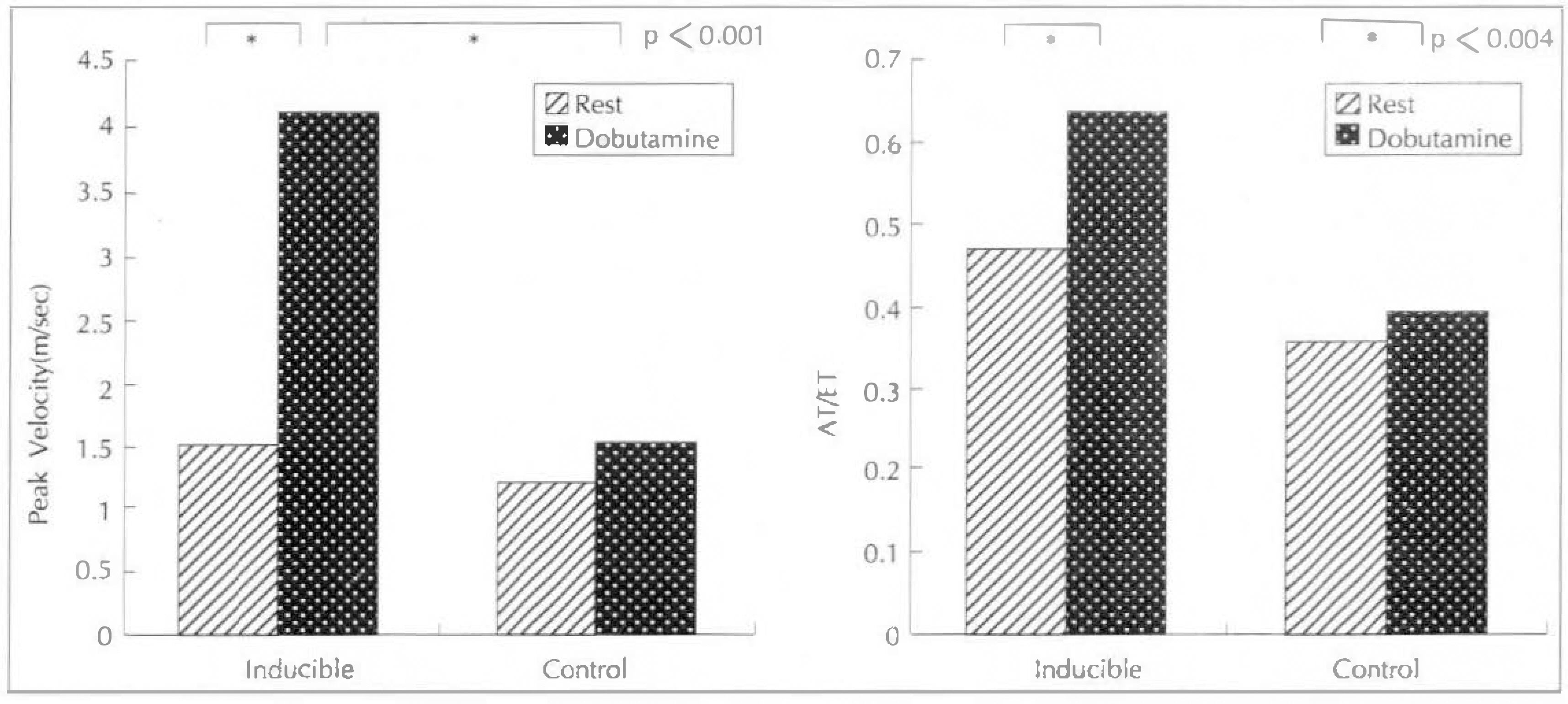
Fig. 2. Change of velocity and AT/ET ratio of outflow tract ject at rest and dobutamine infusion with continous wave Doppler echocardiography.
Reference
-
References
1). Wigle BD, Sasson Z, Henderson MA, Rudy TD, Fulop J, Rakowski H, Williams W. Hypertrophic cardiomyopathy. The importance of the site and extent of hypertrophy. A review. Prog Cardiovasc Dis. 28:1. 1985.2). Marwick TH, Nakatani S, Haluska B, Thomas JD, Lever HM. Provocation of latent left ventricular outflow tract gradients with amyl nitrite and exercise in hypertrophic cardiomyopathy. Am J Cardiol. 75:805. 1995.
Article3). Nakatani S, Lever HM, Marwick TH, Thomas JD. Resting echocardiography identifies hypertrophic cardiomyopathy patients with latent left ventricular outflow obstruction. J Am Coll Cardiol. 25:273A. 1995.4). Rakowski H, Henderson MA, Pollick C. Latent muscular subaortic stenosis. A less severe form of hypertrophic cardiomyopathy. Circulation. 66:118. (abst). 1982.5). Pollick C, Rakowski H, Wigle ED. Muscular subadrtic stenosis. The quantitative relationship between systolic anterior motion and the pressure gradient. Circulation. 69:43. 1984.6). 김권상 정호엔 – 조정휘 검영식 송정상 배종 화 ‘ 페쇄성 비후형 성근증의 Doppler 섬 초음파도 소견. 순환기. 18:647. 1988.7). Klues HG, Leuner C, Kuhn H. Left ventricular outflow tract obstruction in patients with hypertrophic cardiomyopathy. Increase in gradient after exercise. J Am Coll Cardiol. 19:527. 1992.
Article8). Rakowski H, Sasson Z, Wiegle WD. Echocardiographic and Doppler assessment of hypertrophic cardiomyopathy. J Am Soc Echo. 1:31. 1988.
Article9). Yock PG, Hatle L, Popp RL. Pattern and timing of Doppler-detected intracavitary and aortic flow in hypertrophic cardiomyopathy. J Am Coll Cardiol. 8:1047. 1986.10). Schwammenthal E, Schwartzkdpff B, Block M, Johns J, Losse B, Engberding R, Borggrefe M, Breithardt G. Doppler echocardiographic assessment of the pressure gradient during bicycle ergometry in hypertrophic cardiomyopathy. Am J Cardiol. 69:1623. 1992.
Article11). Bryg RJ, Pearson AC, Williams GA, Labovitz AJ. Left ventricular systolic diastolic flow abnormalities determined by Doppler echocardiography in obstructive hypertrophic cardiomyopathy. Am J Cardiol. 59:925. 1987.12). Stewart WJ, Schiavone WA, Salcedo EE, Lever HM, Cosgrove DM, Gill CC. Intraoperative Doppler echdcardiography in hypertrophic cardiomyopathy: correlation with the obstuctive gradient. J Am Coll Cardiol. 10:327. 1987.13). 강흉선 – 조정휘 – 김권삼 ‘ 검병식 · 송정상 · 배종 화. 관동액질환 진단에 있어서 dobutamine 부하 심초음파도 검사의 유용성. 한국심초음파학회지. 1169:1993.14). Movsowitz H, Lampert C, Ioli A, Jacobs LE, Kotler MN. Chest pain in association with dynamic left ventricular outflow obstruction during dobutamine stress testing. J Am Coll Cardiol. 23:142A. 1994.15). Sorrentino MJ, Marcus RH, Lang RM. Left ventricular outflow tract obstruction as a cause for hypotension and symptoms during dobutamine stress echocardiography. Clin. Cardiol. 19:225. 1996.
- Full Text Links
-
- Actions
-
Cited
- CITED
-
- Close
- Share
-
- Similar articles
-
- A case of hypertrophic obstructive cardiomyopathy complicated by infective endocarditis and treated by surgical intervention
- A Case of Regressed Apical Hypertrophic Cardiomyopathy
- Significance of Left Ventricle Chamber Obliteration in Dobutamine Stress Echocardiography
- Usefulness of dobutamine stress echocardiography in the diagnosis of coronary artery disease
- The Usefulness of Dobutamine Stress Echocardiography for Evaluation of Viable Myocardium in Hibernating Myocardium
