Brain Tumor Res Treat.
2018 Apr;6(1):1-7. 10.14791/btrt.2018.6.e4.
Risk of Brain Tumor Induction from Pediatric Head CT Procedures: A Systematic Literature Review
- Affiliations
-
- 1Department of Neurosurgery, David Geffen School of Medicine of the University of California, Los Angeles (UCLA), Los Angeles, CA, USA. iyang@mednet.ucla.edu
- 2Department of Radiological Sciences, Section of Neuroradiology, David Geffen School of Medicine of the University of California, Los Angeles (UCLA), Los Angeles, CA, USA.
- 3Department of Head and Neck Surgery, David Geffen School of Medicine of the University of California, Los Angeles (UCLA), Los Angeles, CA, USA.
- 4Department of Radiation Oncology, David Geffen School of Medicine of the University of California, Los Angeles (UCLA), Los Angeles, CA, USA.
- 5Jonsson Comprehensive Cancer Center, David Geffen School of Medicine of the University of California, Los Angeles (UCLA), Los Angeles, CA, USA.
- 6Los Angeles Biomedical Research Institute, David Geffen School of Medicine of the University of California, Los Angeles (UCLA), Los Angeles, CA, USA.
- 7Harbor-UCLA Medical Center, David Geffen School of Medicine of the University of California, Los Angeles (UCLA), Los Angeles, CA, USA.
- KMID: 2410231
- DOI: http://doi.org/10.14791/btrt.2018.6.e4
Abstract
- Head computed tomography (CT) is instrumental for managing patients of all ages. However, its low dose radiation may pose a low but non-zero risk of tumor induction in pediatric patients. Here, we present a systematic literature review on the estimated incidence of brain tumor induction from head CT exams performed on children and adolescents. MEDLINE was searched using an electronic protocol and bibliographic searches to identify articles related to CT, cancer, and epidemiology or risk assessment. Sixteen studies that predicted or measured head CT-related neoplasm incidence or mortality were identified and reviewed. Epidemiological studies consistently cited increased tumor incidence in pediatric patients (ages 0-18) exposed to head CTs. Excess relative risk of new brain tumor averaged 1.29 (95% confidence interval, 0.66-1.93) for pediatric patients exposed to one or more head CTs. Tumor incidence increased with number of pediatric head CTs in a dose-dependent manner, with measurable excess incidence even after a single scan. Converging evidence from epidemiological studies supported a small excess risk of brain tumor incidence after even a single CT exam in pediatric patients. However, refined epidemiological methods are needed to control for confounding variables that may contribute to reverse causation, such as patients with pre-existing cancer or cancer susceptibility. CT remains an invaluable technology that should be utilized so long as there is clinical indication for the study and the radiation dose is as small as reasonably achievable.
Keyword
MeSH Terms
Figure
-
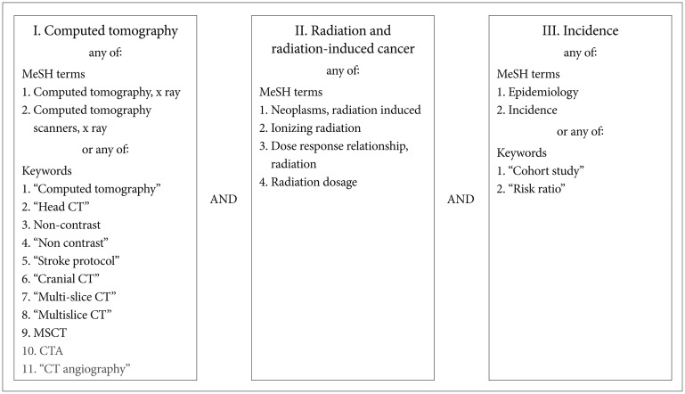
Fig. 1 Protocol used for electronic search of MEDLINE database via PubMed. Identified articles contained at least one relevant keyword or MeSH from each of three major search categories (columns). Grey terms at bottom of left column relate to CT angiography and were subsequently excluded. MeSH, medical subject heading.
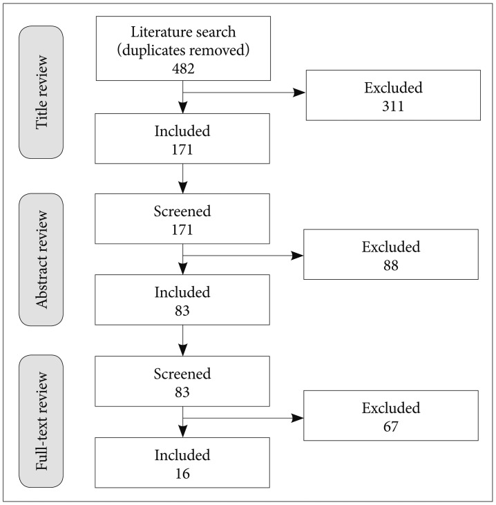
Fig. 2 Summary of article search strategy. Numbers of surviving articles at each stage of screening and review are indicated.
Reference
-
2. Hounsfield GN. Computerized transverse axial scanning (tomography).1. Description of system. Br J Radiol. 1973; 46:1016–1022. PMID: 4757352.3. Ambrose J. Computerized transverse axial scanning (tomography). 2. Clinical application. Br J Radiol. 1973; 46:1023–1047. PMID: 4757353.4. IMV Medical Information Division I. 2016 CT market outlook report. Des Plaines, IL: IMV;2016.5. Brenner DJ, Hall EJ. Computed tomography--an increasing source of radiation exposure. N Engl J Med. 2007; 357:2277–2284. PMID: 18046031.6. Smith-Bindman R, Lipson J, Marcus R, et al. Radiation dose associated with common computed tomography examinations and the associated lifetime attributable risk of cancer. Arch Intern Med. 2009; 169:2078–2086. PMID: 20008690.
Article7. McCollough CH, Bushberg JT, Fletcher JG, Eckel LJ. Answers to common questions about the use and safety of CT scans. Mayo Clin Proc. 2015; 90:1380–1392. PMID: 26434964.
Article8. Friedberg W, Copeland K, Duke FE, O'Brien K 3rd, Darden EB Jr. Radiation exposure during air travel: guidance provided by the Federal Aviation Administration for air carrier crews. Health Phys. 2000; 79:591–595. PMID: 11045535.
Article9. Oksanen PJ. Estimated individual annual cosmic radiation doses for flight crews. Aviat Space Environ Med. 1998; 69:621–625. PMID: 9681366.10. Feng YJ, Chen WR, Sun TP, Duan SY, Jia BS, Zhang HL. Estimated cosmic radiation doses for flight personnel. Space Med Med Eng (Beijing). 2002; 15:265–269. PMID: 12422870.11. The 2007 Recommendations of the International Commission on Radiological Protection. ICRP publication 103. Ann ICRP. 2007; 37:1–332.12. United Nations. Scientific Committee on the Effects of Atomic Radiation. Sources and effects of ionizing radiation: United Nations Scientific Committee on the Effects of Atomic Radiation: UNSCEAR 2000 report to the General Assembly, with scientific annexes. New York: United Nations;2000.13. National Council on Radiation Protection and Measurements. Ionizing radiation exposure of the population of the United States. National Council on Radiation Protection report no. 160. Bethesda (MD): National Council on Radiation Protection and Measurements;2009.14. Mathews JD, Forsythe AV, Brady Z, et al. Cancer risk in 680,000 people exposed to computed tomography scans in childhood or adolescence: data linkage study of 11 million Australians. BMJ. 2013; 346:f2360. PMID: 23694687.15. Schwartz DT. Counter-Point: are we really ordering too many CT scans? West J Emerg Med. 2008; 9:120–122. PMID: 19561720.16. Brenner D, Elliston C, Hall E, Berdon W. Estimated risks of radiation-induced fatal cancer from pediatric CT. AJR Am J Roentgenol. 2001; 176:289–296. PMID: 11159059.
Article17. Tien HC, Tremblay LN, Rizoli SB, et al. Radiation exposure from diagnostic imaging in severely injured trauma patients. J Trauma. 2007; 62:151–156. PMID: 17215747.
Article18. Salibi PN, Agarwal V, Panczykowski DM, et al. Lifetime attributable risk of cancer from CT among patients surviving severe traumatic brain injury. AJR Am J Roentgenol. 2014; 202:397–400. PMID: 24370078.
Article19. Koral K, Blackburn T, Bailey AA, Koral KM, Anderson J. Strengthening the argument for rapid brain MR imaging: estimation of reduction in lifetime attributable risk of developing fatal cancer in children with shunted hydrocephalus by instituting a rapid brain MR imaging protocol in lieu of Head CT. AJNR Am J Neuroradiol. 2012; 33:1851–1854. PMID: 22555583.
Article20. Aw-Zoretic J, Seth D, Katzman G, Sammet S. Estimation of effective dose and lifetime attributable risk from multiple head CT scans in ventriculoperitoneal shunted children. Eur J Radiol. 2014; 83:1920–1924. PMID: 25130177.
Article21. Moher D, Liberati A, Tetzlaff J, Altman DG. PRISMA Group. Preferred reporting items for systematic reviews and meta-analyses: the PRISMA statement. PLoS Med. 2009; 6:e1000097. PMID: 19621072.
Article22. Liberati A, Altman DG, Tetzlaff J, et al. The PRISMA statement for reporting systematic reviews and meta-analyses of studies that evaluate health care interventions: explanation and elaboration. PLoS Med. 2009; 6:e1000100. PMID: 19621070.
Article23. Pflugbeil S, Pflugbeil C, Schmitz-Feuerhake I. Risk estimates for meningiomas and other late effects after diagnostic X-ray exposure of the skull. Radiat Prot Dosimetry. 2011; 147:305–309. PMID: 21831864.
Article24. Chodick G, Ronckers CM, Shalev V, Ron E. Excess lifetime cancer mortality risk attributable to radiation exposure from computed tomography examinations in children. Isr Med Assoc J. 2007; 9:584–587. PMID: 17877063.25. Miglioretti DL, Johnson E, Williams A, et al. The use of computed tomography in pediatrics and the associated radiation exposure and estimated cancer risk. JAMA Pediatr. 2013; 167:700–707. PMID: 23754213.
Article26. Pokora R, Krille L, Dreger S, et al. Computed tomography in Germany. Dtsch Arztebl Int. 2016; 113:721–728. PMID: 27866569.
Article27. Stein SC, Hurst RW, Sonnad SS. Meta-analysis of cranial CT scans in children. A mathematical model to predict radiation-induced tumors. Pediatr Neurosurg. 2008; 44:448–457. PMID: 19018153.28. Brenner DJ, Hall EJ. Cancer risks from CT scans: now we have data, what next? Radiology. 2012; 265:330–331. PMID: 22915598.
Article29. Journy N, Ancelet S, Rehel JL, et al. Predicted cancer risks induced by computed tomography examinations during childhood, by a quantitative risk assessment approach. Radiat Environ Biophys. 2014; 53:39–54. PMID: 24105448.
Article30. Feng ST, Law MW, Huang B, et al. Radiation dose and cancer risk from pediatric CT examinations on 64-slice CT: a phantom study. Eur J Radiol. 2010; 76:e19–e23. PMID: 20363573.
Article31. Pearce MS, Salotti JA, Little MP, et al. Radiation exposure from CT scans in childhood and subsequent risk of leukaemia and brain tumours: a retrospective cohort study. Lancet. 2012; 380:499–505. PMID: 22681860.
Article32. Huang WY, Muo CH, Lin CY, et al. Paediatric head CT scan and subsequent risk of malignancy and benign brain tumour: a nation-wide population-based cohort study. Br J Cancer. 2014; 110:2354–2360. PMID: 24569470.
Article33. Journy N, Rehel JL, Ducou Le Pointe H, et al. Are the studies on cancer risk from CT scans biased by indication? Elements of answer from a large-scale cohort study in France. Br J Cancer. 2015; 112:185–193. PMID: 25314057.
Article34. Krille L, Dreger S, Schindel R, et al. Risk of cancer incidence before the age of 15 years after exposure to ionising radiation from computed tomography: results from a German cohort study. Radiat Environ Biophys. 2015; 54:1–12. PMID: 25567615.35. Berrington de Gonzalez A, Salotti JA, McHugh K, et al. Relationship between paediatric CT scans and subsequent risk of leukaemia and brain tumours: assessment of the impact of underlying conditions. Br J Cancer. 2016; 114:388–394. PMID: 26882064.
Article36. King MA, Kanal KM, Relyea-Chew A, Bittles M, Vavilala MS, Hollingworth W. Radiation exposure from pediatric head CT: a bi-institutional study. Pediatr Radiol. 2009; 39:1059–1065. PMID: 19554322.
Article37. Chen JX, Kachniarz B, Gilani S, Shin JJ. Risk of malignancy associated with head and neck CT in children: a systematic review. Otolaryngol Head Neck Surg. 2014; 151:554–566. PMID: 25052516.38. Pierce DA, Preston DL. Radiation-related cancer risks at low doses among atomic bomb survivors. Radiat Res. 2000; 154:178–186. PMID: 10931690.
Article39. Chomentowski M, Kellerer AM, Pierce DA. Radiation dose dependences in the atomic bomb survivor cancer mortality data: a modelfree visualization. Radiat Res. 2000; 153:289–294. PMID: 10669550.
Article40. Jaffurs D, Denny A. Diagnostic pediatric computed tomographic scans of the head: actual dosage versus estimated risk. Plast Reconstr Surg. 2009; 124:1254–1260. PMID: 19935310.
Article41. Rosen NS. Taking care of children. AJR Am J Roentgenol. 2001; 177:715–717.
Article42. Bertell R, Ehrle LH, Schmitz-Feuerhake I. Pediatric CT research elevates public health concerns: low-dose radiation issues are highly politicized. Int J Health Serv. 2007; 37:419–439. PMID: 17844927.
Article43. Arrangoiz R, Opreanu RC, Mosher BD, Morrison CA, Stevens P, Kepros JP. Reduction of radiation dose in pediatric brain CT is not associated with missed injuries or delayed diagnosis. Am Surg. 2010; 76:1255–1259. PMID: 21140695.
Article44. Pindrik J, Huisman TA, Mahesh M, Tekes A, Ahn ES. Analysis of limited-sequence head computed tomography for children with shunted hydrocephalus: potential to reduce diagnostic radiation exposure. J Neurosurg Pediatr. 2013; 12:491–500. PMID: 24053675.
Article45. Morton RP, Reynolds RM, Ramakrishna R, et al. Low-dose head computed tomography in children: a single institutional experience in pediatric radiation risk reduction: clinical article. J Neurosurg Pediatr. 2013; 12:406–410. PMID: 23971634.
- Full Text Links
-
- Actions
-
Cited
- CITED
-
- Close
- Share
-
- Similar articles
-
- Systematic Review for Remdesivir Use in Pediatric Patients under 3.5 kg with COVID-19
- Is Routine Repeated Head CT Necessary for All Pediatric Traumatic Brain Injury?
- Patients on Anticoagulants after a Head Trauma : Is a Negative Initial CT Scan Enough? Report of a Case of Delayed Subdural Haematoma and Review of the Literature
- Preenhanced computed tomographic findings in brain death
- Risk stratification of intermediate-risk children with minor head injury: a secondary publication translated into Korean
