Comparison of accuracy between panoramic radiography, cone-beam computed tomography, and ultrasonography in detection of foreign bodies in the maxillofacial region: an in vitro study
- Affiliations
-
- 1Dental Implants Research Center, School of Dentistry, Isfahan University of Medical Science, Isfahan, Iran.
- 2Department of Radiology, School of Dentistry, Isfahan University of Medical Science, Isfahan, Iran. maedeh_aminian@yahoo.com
- 3Department of Oral and Maxillo-Facial Surgery, School of Dentistry, Islamic Azad University, Isfahan (Khorasgan) Branch, Isfahan, Iran.
- KMID: 2405372
- DOI: http://doi.org/10.5125/jkaoms.2018.44.1.18
Abstract
OBJECTIVES
Foreign bodies (FBs) account for 3.8% of all pathologies of the head and neck region, and approximately one third of them are missed on initial examination. Thus, FBs represent diagnostic challenges to maxillofacial surgeons, rendering it necessary to employ an appropriate imaging modality in suspected cases.
MATERIALS AND METHODS
In this cross-sectional study, five different materials, including wood, metal, glass, tooth and stone, were prepared in three sizes (0.5, 1, and 2 mm) and placed in three locations (soft tissue, air-filled space and bone surface) within a sheep's head (one day after death) and scanned by panoramic radiography, cone-beam computed tomography (CBCT), and ultrasonography (US) devices. The images were reviewed, and accuracy of the detection modalities was recorded. The data were analyzed statistically using the Kruskal-Wallis, Mann-Whitney U-test, Friedman, Wilcoxon signed-rank and kappa tests (P < 0.05).
RESULTS
CBCT was more accurate in detection of FBs than panoramic radiography and US (P < 0.001). Metal was the most visible FB in all of modalities. US was the most accurate technique for detecting wooden materials, and CBCT was the best modality for detecting all other materials, regardless of size or location (P < 0.05). The detection accuracy of US was greater in soft tissue, while both CBCT and panoramic radiography had minimal accuracy in detection of FBs in soft tissue.
CONCLUSION
CBCT was the most accurate detection modality for all the sizes, locations and compositions of FBs, except for the wooden materials. Therefore, we recommend CBCT as the gold standard of imaging for detecting FBs in the maxillofacial region.
Keyword
MeSH Terms
Figure
-
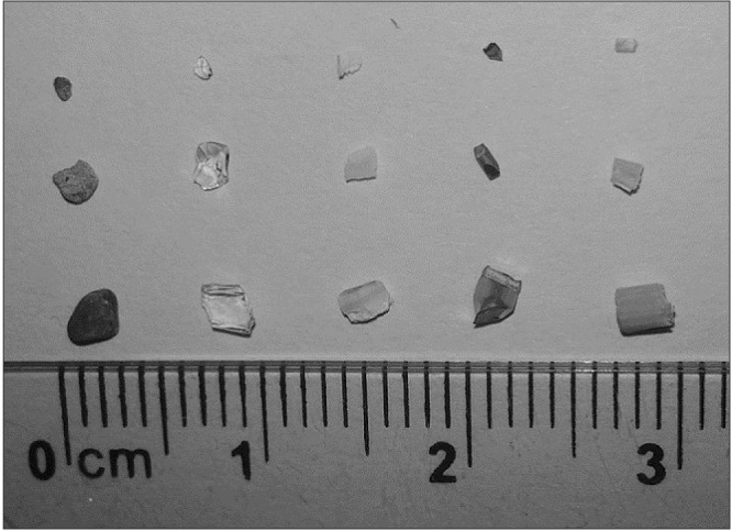
Fig. 1 Materials used as foreign bodies (from left to right: stone, glass, tooth, metal, and wood).
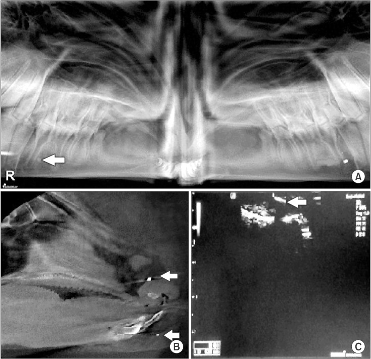
Fig. 2 A. Panoramic radiography view of 2 mm metal (left) and 2 mm stone (right) on bone surface. B. Cone-beam computed tomography sagittal view of 2 mm metal in air-filled space (upper arrow) and 1 mm tooth fragment in soft tissue (lower arrow). C. Ultrasonography view of 2 mm wood in soft tissue (arrow).
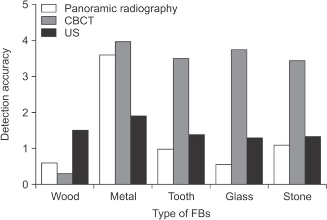
Fig. 3 Detection accuracy of panoramic radiography, cone-beam computed tomography (CBCT) and ultrasonography (US) for different type of foreign bodies (FBs).
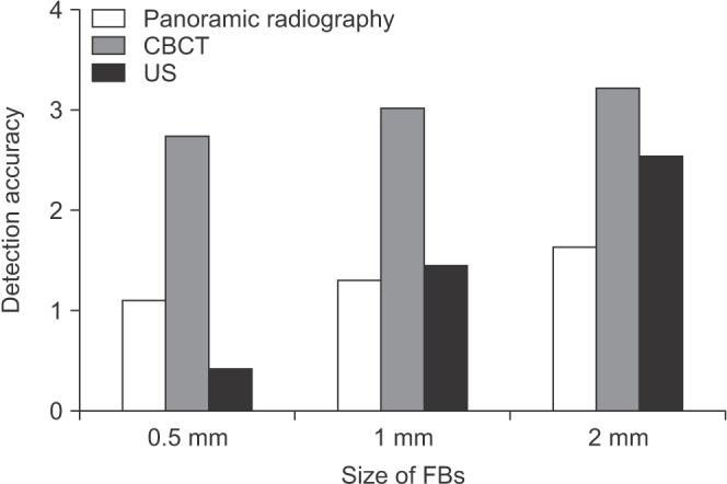
Fig. 4 Detection accuracy of panoramic radiography, cone-beam computed tomography (CBCT) and ultrasonography (US) for different size of foreign bodies (FBs).
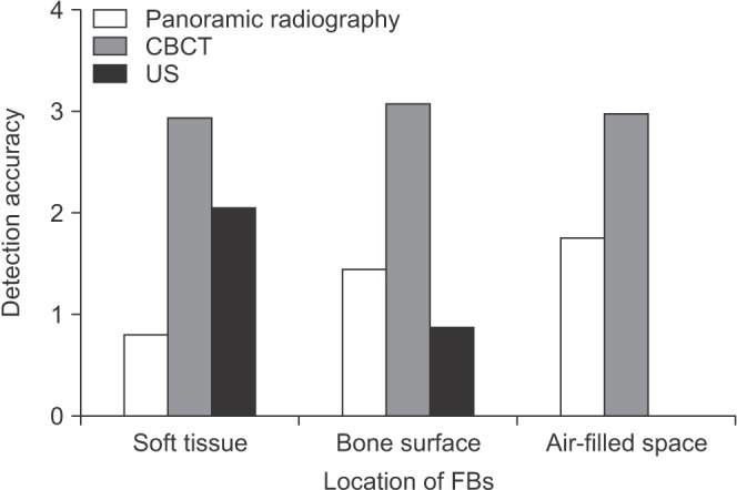
Fig. 5 The accuracy of the panoramic radiography, cone-beam computed tomography (CBCT) and ultrasonography (US) in detection of foreign bodies (FBs) in different locations.
Reference
-
1. Reginelli A, Santagata M, Urraro F, Somma F, Izzo A, Cappabianca S, et al. Foreign bodies in the maxillofacial region: assessment with multidetector computed tomography. Semin Ultrasound CT MR. 2015; 36:2–7. PMID: 25639172.
Article2. Valizadeh S, Pouraliakbar H, Kiani L, Safi Y, Alibakhshi L. Evaluation of visibility of foreign bodies in the maxillofacial region: comparison of computed tomography, cone beam computed tomography, ultrasound and magnetic resonance imaging. Iran J Radiol. 2016; 13:e37265. PMID: 27895878.
Article3. Aras MH, Miloglu O, Barutcugil C, Kantarci M, Ozcan E, Harorli A. Comparison of the sensitivity for detecting foreign bodies among conventional plain radiography, computed tomography and ultrasonography. Dentomaxillofac Radiol. 2010; 39:72–78. PMID: 20100917.
Article4. Omezli MM, Torul D, Sivrikaya EC. The prevalence of foreign bodies in jaw bones on panoramic radiography. Indian J Dent. 2015; 6:185–189. PMID: 26752878.
Article5. Ober CP, Jones JC, Larson MM, Lanz OI, Werre SR. Comparison of ultrasound, computed tomography, and magnetic resonance imaging in detection of acute wooden foreign bodies in the canine manus. Vet Radiol Ultrasound. 2008; 49:411–418. PMID: 18833946.
Article6. Eggers G, Mukhamadiev D, Hassfeld S. Detection of foreign bodies of the head with digital volume tomography. Dentomaxillofac Radiol. 2005; 34:74–79. PMID: 15829688.
Article7. Haghnegahdar A, Shakibafard A, Khosravifard N. Comparison between computed tomography and ultrasonography in detecting foreign bodies regarding their composition and depth: an in vitro study. J Dent (Shiraz). 2016; 17:177–184. PMID: 27602392.8. Oikarinen KS, Nieminen TM, Mäkäräinen H, Pyhtinen J. Visibility of foreign bodies in soft tissue in plain radiographs, computed tomography, magnetic resonance imaging, and ultrasound. An in vitro study. Int J Oral Maxillofac Surg. 1993; 22:119–124. PMID: 8320449.9. Kaviani F, Javad Rashid R, Shahmoradi Z, Gholamian M. Detection of foreign bodies by spiral computed tomography and cone beam computed tomography in maxillofacial regions. J Dent Res Dent Clin Dent Prospects. 2014; 8:166–171. PMID: 25346836.10. Javadrashid R, Fouladi DF, Golamian M, Hajalioghli P, Daghighi MH, Shahmorady Z, et al. Visibility of different foreign bodies in the maxillofacial region using plain radiography, CT, MRI and ultrasonography: an in vitro study. Dentomaxillofac Radiol. 2015; 44:20140229. PMID: 25426703.11. Gomaa M, Abdelaal A. Ultrasonography versus radiography in detection of different foreign bodies in a cadaveric calf thigh specimen. Res J Vet Pract. 2015; 3:83–88.
Article12. Holmes PJ, Miller JR, Gutta R, Louis PJ. Intraoperative imaging techniques: a guide to retrieval of foreign bodies. Oral Surg Oral Med Oral Pathol Oral Radiol Endod. 2005; 100:614–618. PMID: 16243249.
Article13. White SC, Pharaoh MJ. Oral radiology, principles and interpretations. St. Louis: Mosby;2014. p. 111–220.14. Javadrashid R, Golamian M, Shahrzad M, Hajalioghli P, Shahmorady Z, Fouladi DF, et al. Visibility of different intraorbital foreign bodies using plain radiography, computed tomography, magnetic resonance imaging, and cone-beam computed tomography: an in vitro study. Can Assoc Radiol J. 2017; 68:194–201. PMID: 26899378.
Article15. Ginsburg MJ, Ellis GL, Flom LL. Detection of soft-tissue foreign bodies by plain radiography, xerography, computed tomography, and ultrasonography. Ann Emerg Med. 1990; 19:701–703. PMID: 2188542.
Article16. Roobottom CA, Weston MJ. The detection of foreign bodies in soft tissue--comparison of conventional and digital radiography. Clin Radiol. 1994; 49:330–332. PMID: 8013198.
Article17. Ganguly R, Ruprecht A, Vincent S, Hellstein J, Timmons S, Qian F. Accuracy of linear measurement in the Galileos cone beam computed tomography under simulated clinical conditions. Dentomaxillofac Radiol. 2011; 40:299–305. PMID: 21697155.
Article18. Lari SS, Shokri A, Hosseinipanah SM, Rostami S, Sabounchi SS. Comparative sensitivity assessment of cone beam computed tomography and digital radiography for detecting foreign bodies. J Contemp Dent Pract. 2016; 17:224–229. PMID: 27207202.19. Anderson MA, Newmeyer WL 3rd, Kilgore ES Jr. Diagnosis and treatment of retained foreign bodies in the hand. Am J Surg. 1982; 144:63–67. PMID: 7091533.
Article20. Krimmel M, Cornelius CP, Stojadinovic S, Hoffmann J, Reinert S. Wooden foreign bodies in facial injury: a radiological pitfall. Int J Oral Maxillofac Surg. 2001; 30:445–447. PMID: 11720049.
Article21. Specht CS, Varga JH, Jalali MM, Edelstein JP. Orbitocranial wooden foreign body diagnosed by magnetic resonance imaging. Dry wood can be isodense with air and orbital fat by computed tomography. Surv Ophthalmol. 1992; 36:341–344. PMID: 1566235.
Article22. Bushong SC. Radiologic science for technologists: physics, biology, and protection. St. Louis: Elsevier Mosby;2013.
- Full Text Links
-
- Actions
-
Cited
- CITED
-
- Close
- Share
-
- Similar articles
-
- Detection of different foreign bodies in the maxillofacial region with spiral computed tomography and cone-beam computed tomography: An in vitro study
- Comparison of panoramic radiography and cone beam computed tomography for assessing the relationship between the maxillary sinus floor and maxillary molars
- The value of panoramic radiography in assessing maxillary sinus inflammation
- Radiographic evaluation of the course and visibility of the mandibular canal
- Comparison of cone beam computed tomography and conventional panoramic radiography in assessing the topographic relationship between the mandibular canal and impacted third molars
