The Effectiveness and Safety of the Phaco Prechopper Technique Before Lens Phacoemulsification in Cataract Surgery
- Affiliations
-
- 1Department of Ophthalmology and Visual Science, Kangnam St. Mary's Hospital, College of Medicine, The Catholic University of Korea, Seoul, Korea. ckjoo@catholic.ac.kr
Abstract
-
PURPOSE: To compare the effectiveness and safety of the phaco prechopper technique dividing lenses into 2 or 4 pieces before phacoemulsification with the effectiveness and safety of conventional phacoemulsification during cataract surgery.
METHODS
This study included 360 eyes, which were divided into 4 groups according to nuclear opacity each group was subdivided into 3 groups according to the lens extraction technique (control, dividing the lens into two pieces using a phaco prechopper, and dividing it into four pieces) (Table 1). The following parameters were compared between the groups that had the same degree of nuclear opacity: changes in the endothelial cell count and hexagonality 2 months postoperatively, phaco time as well as the total phaco energy used for the operation, and the complication rates.
RESULTS
There were no intra- and post-operative complications in any group. The endothelial cell loss rate was significantly less in nuclear opacity groups 2 and 3 with the prechopper technique than in the control group. Hexagonality was significantly higher in the nuclear opacity group 3 with the prechopper technique than in the control 2 months after the operation. Phaco time was significantly less in the nuclear opacity groups 2 and 3 with the prechopper technique than in the control. Total phaco energy was significantly less in the nuclear opacity groups 2, 3, and 4 with the prechopper technique than in the control.
CONCLUSIONS
Phacoemulsification using the prechopper technique is safe, resulting in significantly less endothelial cell damage, and requires less phaco time and less total phaco energy compared to those of the conventional phacoemulsification technique.
Keyword
Figure
-

Figure 1. Comparison of corneal endothelial cell loss rate (%) among various phacoemulsification techniques prior to and two months after cataract operation. *1 P=0.047: Multiple comparisons (Tukey HSD and Duncan test) for NO2, *2 P=0.048: Multiple comparisons (Tukey HSD and Duncan test) for NO2 , **1 P=0.04 : Multiple comparisons (Tukey HSD and Duncan test) for NO3, **2 P=0.04: Multiple comparisons (Tukey HSD and Duncan test) for NO3 groups (Control group: Groups not using phaco prechopper).
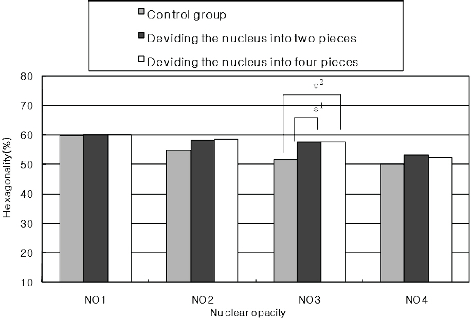
Figure 3 . Comparison of phaco time (sec) between subgroups using different phacoemulsification techniques. *1 P=0.04: Multiple comparisons (Tukey HSD and Duncan test) for NO2, *2 P=0.04: Multiple comparisons (Tukey HSD and Duncan test) for NO2, **1 P=0.04: Multiple comparisons (Tukey HSD and Duncan test) for NO3, **2 P=0.04: Multiple comparisons (Tukey HSD and Duncan test) for NO3 groups (Control group: Groups not using phaco prechopper).

Figure 2. Comparison of corneal endothelial cell hexagonality (%) two months after cataract operation among various phacoemulsification techniques. *1 P=0.03: Multiple comparisons (Tukey HSD and Duncan test) for NO3, *2 P=0.04: Multiple comparisons (Tukey HSD and Duncan test) for NO3 (Control group: Groups not using phaco pre- chopper).

Figure 4. Comparison of absolute phaco time (Absolute phaco time (%·sec)) delivered between subgroups using different phacoemulsification techniques. The absolute phaco time is calculated by multiplying the phaco power and time. *1 P=0.04: Multiple comparisons (Tukey HSD and Duncan test) for NO2, *2 P=0.04: Multiple comparisons (Tukey HSD and Duncan test) for NO2 , **1 P=0.04: Multiple comparisons (Tukey HSD and Duncan test) for NO3, **2 P=0.03: Multiple comparisons (Tukey HSD and Duncan test) for NO3 groups, ***1 P=0.04: Multiple comparisons (Tukey HSD and Duncan test) for NO3, ***2 P=0.04: Multiple comparisons (Tukey HSD and Duncan test) for NO4 groups (Control group: Groups not using phaco prechopper).
Reference
-
References
1. Kim YH, Joo CK. Phacoemulsification by Phaco-drill Method with Microseal Phaco Tip. J Korean Ophthalmol Soc. 1997; 38:1566–71.2. Kim YH, Joo CK. Short-term Change of Corneal Endothelium after Phacoemulsification using Phaco-drill Technique. J Korean Ophthalmol Soc. 1999; 40:81–7.3. Akahoshi T. Phaco prechop: manual nucleofracture prior to phacoemulsification. Operative Techniques in Cataract and Refractive Surgery. 1998; 11:69–91.4. Park YJ, An GJ, Woo CH. Corneal Endothelial Cell Change after Small Incision Cataract Surgery J Korean Ophthalmol Soc. 1998; 39:487–94.5. Polack M, Sugar A. The phacoemulsification procedure Ⅲ. Corneal complications. Invest Ophthalmol Vis Sci. 1977; 16:39–46.6. Gwin RM, Warren JK, Samuelson DA, Gum GG. Effects of phacoemulsification and extracapsular lens removal on corneal thickness and endothelialcell density in the dog. Invest Ophthalmol Vis Sci. 1983; 24:227–36.7. Strobel J, Jacobi KW. Phacoemulsification and planed ECCE: Intraoperative differences in intraocular heating. Eur J implant Ref Surg. 1991; 3:135–8.8. Behndig A, Lundberg B. Transient corneal edema after phacoemulsification : comparison of 3 viscoelstic regimens. J Cataract Refract Surg. 2002; 28:1551–6.9. Davis EA, Lindstorm RL. Corneal thickness and visual acuity after phacoemulsification with 3 viscoelastic materials. J Cataract Refract Surg. 2000; 26:1505–9.
Article10. Arshinoff SA. Dispersive-cohesive viscoeastic soft shell technique. J Cataract Refract Surg. 1999; 25:167–73.11. Arshinoff SA. Using BSS with viscoadaptives in the ultimate soft-shell technique. J Cataract Refract Surg. 2002; 28:1509–14.
Article12. Pirazzoli G, D'Eliseo D, Ziosi M, Acciarri R. Effects of phacoemulsification time on the corneal endothelium using phacofracture and phaco chop techniques. J Cataract Refract Surg. 1996; 22:967–9.
Article13. Joo CK, Kim YH. Phacoemulsification with a bevel-down phaco tip: phaco-drill. J Cataract Refract Surg. 1997; 23:1149–52.
Article14. Fine IH, Packer M, Hoffman RS. Use of power modulations in phacoemulsification; choo-choo chop and flip phacoemulsi- fication. J Cataract Refract Surg. 2001; 27:188–97.15. Badoza D, Fernández Mendy J, Ganly M. Phacoemulsification using the burst mode. J Cataract Refract Surg. 2003; 29:1101–5.
Article16. Fine IH, Packer M, Hoffman RS. New phacoemulsification technologies. J Cataract Refract Surg. 2002; 28:1054–60.
Article17. Durán S, Zati M. Erbium: YAG laser emulsification of the cataractous lens. J Cataract Refract Surg. 2001; 27:1025–32.18. Doughty MJ. Toward a quantitative analysis of corneal endothelial cell morphology: a review of technique and their application. Optom Vis Sci. 1989; 66:626–42.19. Matsuda M, Suda T, Manabe R. Serial alterations in endothelial cell shape and pattern after intraocular surgery. Am J Ophthalmol. 1984; 98:313–9.
Article20. Kok JH, Dunnebier EA, Nieurwendaal CP, Kijlstra A. Polymegethism of the corneal endothelium in an eye with long-standing ptosis. Arch Ophthalmol. 1992; 110:1529–30.
Article21. Díaz-Valle D, Benítez Del Castillo Sanchez JM, Toledano N. . Endothelial morphological and functional evaluation after cataract surgery. Eur J Ophthalmol. 1996; 6:242–5.
Article22. Kraff MC, Sanders DR, Lieberman HL. Specular microscopy in cataract and intraocular lens patients. Arch Ophthalmol. 1980; 98:1782–4.
Article23. Mishima S. Clinical investigations on the corneal endothelium -XXXVIII Edward Jackson Memorial Lecture. Am J Ophthalmol. 1982; 93:1–29.24. Yee RW, Geroski D, Matsuda M. . Correlation of corneal endothelial pump site density, barrier function and morphology in wound repair. Invest Ophthalmol Vis Sci. 1985; 26:1191–201.25. Walkow T, Anders N, Klebe S. Endothelial cell loss after phacoemulsification: relationto preoperative and intraoperative parameters. J Cataract Refract Surg. 2000; 26:727–32.26. Hayashi K, Hayashi H, Nakao F, Hayashi F. Risk factors for corneal endothelial injury during phacoemulsification. J Cataract Refract Surg. 1996; 22:1079–84.
Article
- Full Text Links
-
- Actions
-
Cited
- CITED
-
- Close
- Share
-
- Similar articles
-
- Sutureless Small Incision ECCE with Phaco Prechopper
- Phacoemulsification by Phaco-Drill Method with Microseal Phaco Tip
- Comparison of Surgical Parameters and Outcomes According to the Phacoemulsification Technique
- Clinical Comparison of Crown-shaped Phaco Tip with Conventional Phaco Tip
- Comparison of the Phaco Chop and the Mini Chop Methods in Cataract Surgery
