Differences in the mandibular premolar positions in Angle Class I subjects with different vertical facial types: A cone-beam computed tomography study
- Affiliations
-
- 1Department of Stomatology, Ministry of Education Key Laboratory of Child Development and Disorders, Children's Hospital of Chongqing Medical University, Chongqing, China.
- 2Department of Orthodontics, College of Stomatology, Chongqing Medical University, Chongqing, China. yanjiebai@aol.com
- 3Chongqing Key Laboratory for Oral Diseases and Biomedical Sciences, Chongqing Medical University, Chongqing, China.
- 4Department of Otorhinolaryngology, Children's Hospital of Chongqing Medical University, Chongqing, China.
- KMID: 2400576
- DOI: http://doi.org/10.4041/kjod.2015.45.4.180
Abstract
OBJECTIVE
To compare the positions of the mandibular premolars in Angle Class I subjects according to vertical facial type. The results will provide a theoretical basis for predicting effective tooth movement in orthodontic treatment.
METHODS
Cephalometric parameters were determined using cone-beam computed tomography in 120 Angle Class I subjects. Subjects were categorized as short, normal, and long face types according to the Frankfort mandibular angle. Parameters indicating the position of the mandibular right premolars and the mandible were also measured.
RESULTS
The angle between the mandibular first premolar axis and buccal cortex, the distance between the root apex and buccal cortex, angle of vestibularization, arc of vestibularization, and root apex maximum movable distance were significantly greater in the short face type than in the long and norm face types. The angle between the mandibular second premolar axis and buccal cortex, the distance from root apex to buccal cortex, and the arc of vestibularization were significantly greater in the short face type than in the normal face type.
CONCLUSIONS
There are significant differences in the mandibular premolar positions in Class I subjects according to vertical facial type.
MeSH Terms
Figure
-
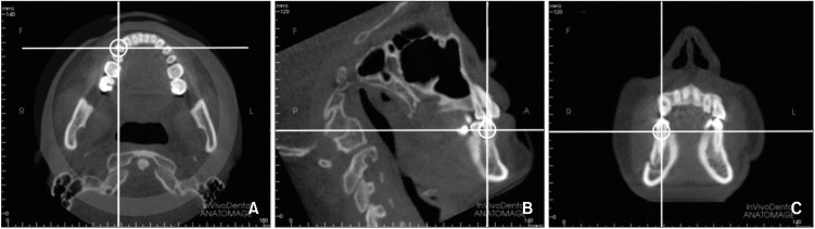
Figure 1 The cone-beam computed tomography image of the craniofacial region using InvivoDental 5.1 software. The transverse (A), sagittal (B), and coronal planes (C) are indicated.
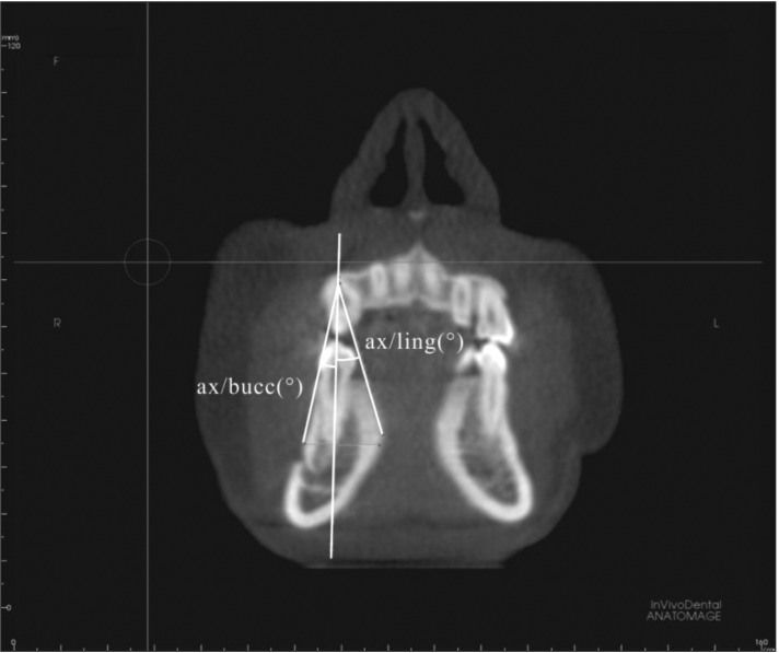
Figure 2 Angle between the axis and the buccal cortex (Premo ax/bucc cort), and angle between the axis and the lingual cortex (Premo ax/ling cort). A representative cone-beam computed tomography image of the mandible through the coronal plane is shown.
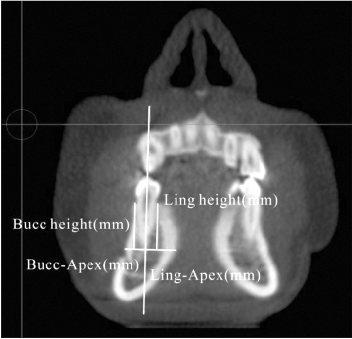
Figure 3 Alveolar thickness and height measurements. A representative cone-beam computed tomography image of the mandible through the coronal plane is shown. Bucc, Buccal; Ling, lingual.
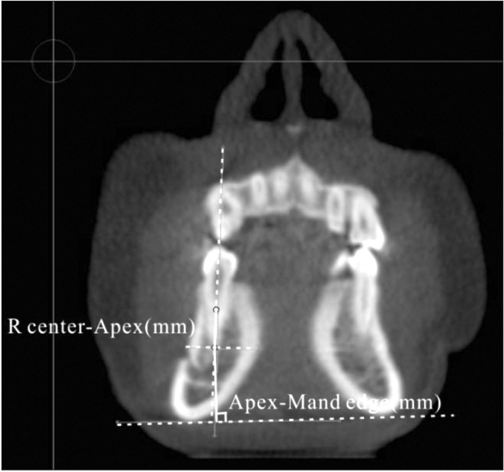
Figure 4 Distance from the resistance center of the mandibular premolar to its apex (R center-Apex) , and distance from the apex to the mandible edge (Apex-Mand edge). A representative cone-beam computed tomography image of the mandible through the coronal plane is shown.
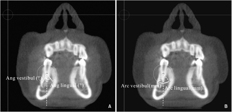
Figure 5 Parameters indicating the range in tooth movement. A, Measurement of the vestibularization and lingualization angles (Ang vestibule and Ang lingual). B, Measurement of the vestibularization and lingualization arcs (Arc vestibule and Arc lingual). A representative cone-beam computed tomography image of the mandible through the coronal plane is shown.
Reference
-
1. Ingervall B, Thilander B. Relation between facial morphology and activity of the masticatory muscles. J Oral Rehabil. 1974; 1:131–147. PMID: 4525024.
Article2. Bassil-Nassif N, Bouserhal J, Garcia R. Facial volumes and vertical facial type: a three dimensional comparative study. Orthod Fr. 2010; 81:127–137. PMID: 20519108.3. Tsunori M, Mashita M, Kasai K. Relationship between facial types and tooth and bone characteristics of the mandible obtained by CT scanning. Angle Orthod. 1998; 68:557–562. PMID: 9851354.4. Gracco A, Lombardo L, Mancuso G, Gravina V, Siciliani G. Upper incisor position and bony support in untreated patients as seen on CBCT. Angle Orthod. 2009; 79:692–702. PMID: 19537878.
Article5. Yamada C, Kitai N, Kakimoto N, Murakami S, Furukawa S, Takada K. Spatial relationships between the mandibular central incisor and associated alveolar bone in adults with mandibular prognathism. Angle Orthod. 2007; 77:766–772. PMID: 17685772.
Article6. Handelman CS. The anterior alveolus: its importance in limiting orthodontic treatment and its influence on the occurrence of iatrogenic sequelae. Angle Orthod. 1996; 66:95–109. discussion 109-10PMID: 8712499.7. Masumoto T, Hayashi I, Kawamura A, Tanaka K, Kasai K. Relationships among facial type, buccolingual molar inclination, and cortical bone thickness of the mandible. Eur J Orthod. 2001; 23:15–23. PMID: 11296507.
Article8. Broadbent BH. A new x-ray technique and its application to orthodontia. Angle Orthod. 1931; 1:45–66.9. Scarfe WC, Farman AG, Sukovic P. Clinical applications of cone-beam computed tomography in dental practice. J Can Dent Assoc. 2006; 72:75–80. PMID: 16480609.10. NA B, XU T, Lin J. Vertical changes during orthodontic treatment for class II division 1 female. Chinese J Orthod. 2006; 13:23–26.11. Timock AM, Cook V, McDonald T, Leo MC, Crowe J, Benninger BL, et al. Accuracy and reliability of buccal bone height and thickness measurements from cone-beam computed tomography imaging. Am J Orthod Dentofacial Orthop. 2011; 140:734–744. PMID: 22051495.
Article12. Sherrard JF, Rossouw PE, Benson BW, Carrillo R, Buschang PH. Accuracy and reliability of tooth and root lengths measured on cone-beam computed tomographs. Am J Orthod Dentofacial Orthop. 2010; 137(4 Suppl):S100–S108. PMID: 20381750.
Article13. Tarazona B, Llamas JM, Cibrian R, Gandia JL, Paredes V. A comparison between dental measurements taken from CBCT models and those taken from a digital method. Eur J Orthod. 2013; 35:1–6. PMID: 21427171.
Article14. Ludlow JB, Gubler M, Cevidanes L, Mol A. Precision of cephalometric landmark identification: cone-beam computed tomography vs conventional cephalometric views. Am J Orthod Dentofacial Orthop. 2009; 136:312.e1–312.10. discussion 312-3PMID: 19732656.
Article15. Hassan B, van der Stelt P, Sanderink G. Accuracy of three-dimensional measurements obtained from cone beam computed tomography surface-rendered images for cephalometric analysis: influence of patient scanning position. Eur J Orthod. 2009; 31:129–134. PMID: 19106265.
Article16. Thilander B, Nyman S, Karring T, Magnusson I. Bone regeneration in alveolar bone dehiscences related to orthodontic tooth movements. Eur J Orthod. 1983; 5:105–114. PMID: 6574916.
Article17. Steiner GG, Pearson JK, Ainamo J. Changes of the marginal periodontium as a result of labial tooth movement in monkeys. J Periodontol. 1981; 52:314–320. PMID: 6943326.
Article18. Kaley J, Phillips C. Factors related to root resorption in edgewise practice. Angle Orthod. 1991; 61:125–132. PMID: 2064070.19. Edwards JG. A study of the anterior portion of the palate as it relates to orthodontic therapy. Am J Orthod. 1976; 69:249–273. PMID: 1062165.
Article20. Sarikaya S, Haydar B, Ciğer S, Ariyürek M. Changes in alveolar bone thickness due to retraction of anterior teeth. Am J Orthod Dentofacial Orthop. 2002; 122:15–26. PMID: 12142888.
Article21. Ten Hoeve A, Mulie RM. The effect of anteropostero incisor repositioning on the palatal cortex as studied with laminagraphy. J Clin Orthod. 1976; 10:804–822. PMID: 1069732.22. Andrews LF. The six keys to normal occlusion. Am J Orthod. 1972; 62:296–309. PMID: 4505873.
Article23. Linge BO, Linge L. Apical root resorption in upper anterior teeth. Eur J Orthod. 1983; 5:173–183. PMID: 6578039.
Article24. Darendeliler MA, Kharbanda OP, Chan EK, Srivicharnkul P, Rex T, Swain MV, et al. Root resorption and its association with alterations in physical properties, mineral contents and resorption craters in human premolars following application of light and heavy controlled orthodontic forces. Orthod Craniofac Res. 2004; 7:79–97. PMID: 15180087.
Article25. Yoshida N, Jost-Brinkmann PG, Koga Y, Mimaki N, Kobayashi K. Experimental evaluation of initial tooth displacement, center of resistance, and center of rotation under the influence of an orthodontic force. Am J Orthod Dentofacial Orthop. 2001; 120:190–197. PMID: 11500662.
Article26. Tanne K, Nagataki T, Inoue Y, Sakuda M, Burstone CJ. Patterns of initial tooth displacements associated with various root lengths and alveolar bone heights. Am J Orthod Dentofacial Orthop. 1991; 100:66–71. PMID: 2069150.
Article27. Choy K, Pae EK, Park Y, Kim KH, Burstone CJ. Effect of root and bone morphology on the stress distribution in the periodontal ligament. Am J Orthod Dentofacial Orthop. 2000; 117:98–105. PMID: 10629526.
Article28. Proffit WR, Fields HW, Nixon WL. Occlusal forces in normal- and long-face adults. J Dent Res. 1983; 62:566–570. PMID: 6573373.
Article
- Full Text Links
-
- Actions
-
Cited
- CITED
-
- Close
- Share
-
- Similar articles
-
- Differences in facial soft tissue deviations in Class III patients with different types of mandibular asymmetry: A cone-beam computed tomography study
- Location and shape of the mandibular lingula: Comparison of skeletal class I and class III patients using panoramic radiography and cone-beam computed tomography
- Evaluation of buccolingual molar inclinations among different vertical facial types
- Three-dimensional analysis of dental decompensation for skeletal Class III malocclusion on the basis of vertical skeletal patterns obtained using cone-beam computed tomography
- Differences in positions of cone-beam computed tomography landmarks in patients with skeletal Class III facial asymmetry according to midsagittal planes
