Pituicytoma with Significant Tumor Vascularity Mimicking Pituitary Macroadenoma
- Affiliations
-
- 1Department of Neurosurgery, Pusan National University School of Medicine, Pusan National University Hospital, Busan, Korea. neurocha@hanmail.net
- 2Department of Pathology, Seoul National University College of Medicine, Seoul National University Hospital, Seoul, Korea.
- KMID: 2396456
- DOI: http://doi.org/10.14791/btrt.2017.5.2.110
Abstract
- A 19-year-old man presented with bitemporal hemianopsia and was found to have a large sellar and suprasellar tumor, resembling a pituitary macroadenoma. Emergency transsphenoidal approach was attempted because of rapid visual deterioration with headache. However, the approach was complicated and stopped by uncontrolled hemorrhage from the tumor. After conventional cerebral angiography and recognition of an unusual pathology, transcranial approach was achieved to prevent permanent visual loss. The final pathological diagnosis was pituicytoma with epithelioid features. Pituicytoma is a rare low-grade tumor (WHO Grade I) of pituicytes involving the sellar and suprasellar region, and originating from special glial cells of the neurohypophysis. Because of the high vascularity, the firm consistency, and invasion to surrounding neurovascular structures, a pituicytoma should be included in the differential diagnosis of a mass in the sellar and suprasellar area if the tumor shows high enhancement with vascular components. We report a case of rare pituicytoma mimicking a pituitary macroadenoma with massive hemorrhage to disturb surgery.
Keyword
MeSH Terms
Figure
-

Fig. 1 Preoperative magnetic resonance imaging. T1-weighted axial image (A) showing slightly hyperintense sellar mass, T2-weighted axial image (B) showing isointense sellar mass, and T1-weighted gadolinium enhanced coronal (C) and sagittal (D) images demonstrating marked enhancement of sellar-suprasellar mass with involvement of the optic nerves and chiasm.

Fig. 2 Angiograms of right (A) and left (B) internal carotid arteries showing significant tumor blush in the arterial phases by multiple small feeders from both internal carotid arteries.
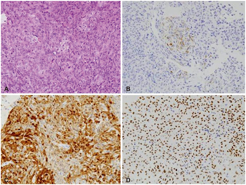
Fig. 3 Histopathological findings of the pituicytoma. A: The tumor cells have moderate to abundant amount of pale eosinophilic cytoplasm and ovoid nuclei with small nucleoli. Necrosis is not identified (hematoxylin and eosin staining, ×400). B: Immunohistochemistry for glial fibrillary acidic protein shows focal positivity (×400). C: The tumor cells are positive for S100 protein (×400). D: The tumor cells show positive staining for thyroid transcription factor 1 (×400).

Fig. 4 Postoperative magnetic resonance imaging after transcranial approach. T1-weighted gadolinium enhanced coronal (A) and sagittal (B) images show total removal of the suprasellar tumor that compressed the optic chiasm and remnants attached to both cavernous sinuses.
Reference
-
1. Louis DN, Ohgaki H, Wiestler OD, et al. The 2007 WHO classification of tumours of the central nervous system. Acta Neuropathol. 2007; 114:97–109.
Article2. Liss L, Kahn EA. Pituicytoma, tumor of the sella turcica; a clinicopathological study. J Neurosurg. 1958; 15:481–488.3. Brat DJ, Scheithauer BW, Staugaitis SM, Holtzman RN, Morgello S, Burger PC. Pituicytoma: a distinctive low-grade glioma of the neurohypophysis. Am J Surg Pathol. 2000; 24:362–368.4. Lee EB, Tihan T, Scheithauer BW, Zhang PJ, Gonatas NK. Thyroid transcription factor 1 expression in sellar tumors: a histogenetic marker. J Neuropathol Exp Neurol. 2009; 68:482–488.
Article5. Bara D, Lantos P. Two rare forms of tumour in the hypothalamo-hypophysial system. Infundibular choristoma and glioblastoma infiltrating the pituitary. Acta Morphol Acad Sci Hung. 1968; 16:243–250.6. Wolfe SQ, Bruce J, Morcos JJ. Pituicytoma: case report. Neurosurgery. 2008; 63:E173–E174. discussion E174.7. Secci F, Merciadri P, Rossi DC, D'Andrea A, Zona G. Pituicytomas: radiological findings, clinical behavior and surgical management. Acta Neurochir (Wien). 2012; 154:649–657.
Article8. Brat DJ, Scheithauer BW, Fuller GN, Tihan T. Newly codified glial neoplasms of the 2007 WHO Classification of Tumours of the Central Nervous System: angiocentric glioma, pilomyxoid astrocytoma and pituicytoma. Brain Pathol. 2007; 17:319–324.
Article9. Gibbs WN, Monuki ES, Linskey ME, Hasso AN. Pituicytoma: diagnostic features on selective carotid angiography and MR imaging. AJNR Am J Neuroradiol. 2006; 27:1639–1642.10. Katsuta T, Inoue T, Nakagaki H, Takeshita M, Morimoto K, Iwaki T. Distinctions between pituicytoma and ordinary pilocytic astrocytoma. Case report. J Neurosurg. 2003; 98:404–406.11. Miki Y, Matsuo M, Nishizawa S, et al. Pituitary adenomas and normal pituitary tissue: enhancement patterns on gadopentetate-enhanced MR imaging. Radiology. 1990; 177:35–38.
Article12. Tian Y, Yue S, Jia G, Zhang Y. Childhood giant pituicytoma: a report and review of the literature. Clin Neurol Neurosurg. 2013; 115:1943–1950.
Article13. Furtado SV, Ghosal N, Venkatesh PK, Gupta K, Hegde AS. Diagnostic and clinical implications of pituicytoma. J Clin Neurosci. 2010; 17:938–943.
Article14. Orrego JJ. Pituicytoma and isolated ACTH deficiency. Pituitary. 2009; 12:371–372.
Article15. Phillips JJ, Misra A, Feuerstein BG, Kunwar S, Tihan T. Pituicytoma:characterization of a unique neoplasm by histology, immunohistochemistry, ultrastructure, and array-based comparative genomic hybridization. Arch Pathol Lab Med. 2010; 134:1063–1069.
Article16. Kim YG, Park YS. Second-stage transsphenoidal approach (TSA) for highly vascular pituicytomas in children. Childs Nerv Syst. 2015; 31:985–989.
Article17. Feng M, Carmichael JD, Bonert V, Bannykh S, Mamelak AN. Surgical management of pituicytomas: case series and comprehensive literature review. Pituitary. 2014; 17:399–413.
Article
- Full Text Links
-
- Actions
-
Cited
- CITED
-
- Close
- Share
-
- Similar articles
-
- Pituitary Apoplexy due to Pituitary Adenoma Infarction
- Pituicytoma: Case Report
- Intrasellar Cavernous Hemangioma
- Comparison of Anterior Pituitary Function between Patients with GH-secreting Macroadenoma and those with Nonfunctioning Macroadenoma
- Two Cases of Cushing's Disease Due to Large Pituitary ACTH Secreting Tumor
