Borderline Phyllodes Tumor with an Incidental Invasive Tubular Carcinoma and Lobular Carcinoma In Situ Component: A Case Report
- Affiliations
-
- 1Department of Radiation Oncology, Juravinski Cancer Centre, McMaster University, Hamilton, Ontario, Canada.
- 2Department of Surgery, Juravinski Cancer Centre, McMaster University, Hamilton, Ontario, Canada.
- 3Department of Pathology, Juravinski Hospital, McMaster University, Hamilton, Ontario, Canada.
- 4Department of Anatomical Pathology, McMaster University Medical Centre, Hamilton, Ontario, Canada. tangsh@hhsc.ca
Abstract
- Phyllodes tumors are an infrequent breast tumor presentation. A phyllodes tumor with a synchronous invasive ductal carcinoma is rarely described and has never been reported with lobular carcinoma in situ component. A 53-year-old female presented with a nine-year history of twice core biopsy proven fibroadenoma. After an increase in the tumor's growth velocity it was decided upon to undergo an excisional biopsy. Microscopic examination of the well-circumscribed pale-tan mass found focal areas of leaf like architecture with variable number of mitoses present, representing a phyllodes tumor of borderline malignant potential. Incidentally, at one edge of the mass was found a tubular carcinoma and lobular carcinoma in situ components. Thorough, routine follow-up of patients with biopsy proven benign breast masses is important to finding a masked malignant component.
MeSH Terms
Figure
-
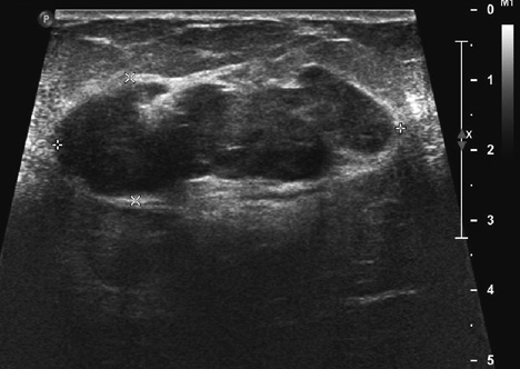
Figure 1 Ultrasound (US) of right breast multilobulated, solid mass measured 4.91×1.76 cm. Mildly heterogenous, paralleling the skin surface. In 2001 the tumor's longest US dimension was 2.4 cm.
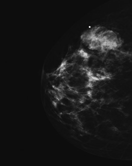
Figure 2 Mammography of right breast, craniocaudal view, showed a circumscribed, multilobulated solid mass.
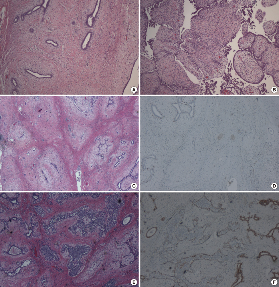
Figure 3 (A) The mass revealed fibroadenomatous change (H&E stain, ×40). (B) Leaf-like structures of fibroepithelial lesion showed a cellular stroma with cytological atypia and increased mitosis (H&E stain, ×40). (C) Invasive carcinoma cells formed small tubules within the stroma (H&E stain, ×20). (D) Tubules of carcinoma cells were immunohistochemically negative for myoepithelial marker, p63 (IHC, ×40). (E) Lobular carcinoma in situ was noted within the glandular component (H&E stain, ×40). (F) Lobular carcinoma in situ was immunohistochemically negative for E-cadherin (×40).
Reference
-
1. Rosen PP, Oberman HA. Rosai J, Sobin LH, editors. Cystosarcoma phyllodes. Atlas of Tumor Pathology. Tumors of the Mammary Gland. 1993. 3rd series. Washington, DC: Armed Forces Institute of Pathology;101–114.2. Ozzello L, Gump FE. The management of patients with carcinomas in fibroadenomatous tumors of the breast. Surg Gynecol Obstet. 1985. 160:99–104.3. Parfitt JR, Armstrong C, O'malley F, Ross J, Tuck AB. In-situ and invasive carcinoma within a phyllodes tumor associated with lymph node metastases. World J Surg Oncol. 2004. 2:46.4. Kodama T, Kameyama K, Mukai M, Sugiura H, Ikeda T, Okada Y. Invasive lobular carcinoma arising in phyllodes tumor of the breast. Virchows Arch. 2003. 442:614–616.
Article5. Macher-Goeppinger S, Marme F, Goeppert B, Penzel R, Schirmacher P, Sinn HP, et al. Invasive ductal breast cancer within a malignant phyllodes tumor: case report and assessment of clonality. Hum Pathol. 2010. 41:293–296.
Article6. Sugie T, Takeuchi E, Kunishima F, Yotsumoto F, Kono Y. A case of ductal carcinoma with squamous differentiation in malignant phyllodes tumor. Breast Cancer. 2007. 14:327–332.
Article7. de Rosa G, Ferrara G, Goglia P, Ghicas C, Zeppa P. In situ and microinvasive carcinoma with squamoid differentiation arising in a phyllodes tumor: report of a case. Tumori. 1989. 75:514–517.
Article8. Reinfuss M, Mituś J, Duda K, Stelmach A, Ryś J, Smolak K. The treatment and prognosis of patients with phyllodes tumor of the breast: an analysis of 170 cases. Cancer. 1996. 77:910–916.
Article9. Barth RJ Jr. Histologic features predict local recurrence after breast conserving therapy of phyllodes tumors. Breast Cancer Res Treat. 1999. 57:291–295.
Article10. Christensen L, Nielsen M, Madsen PM. Cystosarcoma phyllodes. A review of 19 cases with emphasis on the occurrence of associated breast carcinoma. Acta Pathol Microbiol Immunol Scand A. 1986. 94:35–41.11. Yilmaz E, Sal S, Lebe B. Differentiation of phyllodes tumors versus fibroadenomas. Acta Radiol. 2002. 43:34–39.
Article12. Bode MK, Rissanen T, Apaja-Sarkkinen M. Ultrasonography and core needle biopsy in the differential diagnosis of fibroadenoma and tumor phyllodes. Acta Radiol. 2007. 48:708–713.
Article
- Full Text Links
-
- Actions
-
Cited
- CITED
-
- Close
- Share
-
- Similar articles
-
- Multi-Focal Lobular Carcinoma In Situ Arising in Benign Phyllodes Tumor: A Case Report
- Invasive Lobular Carcinoma of the Breast Associated with Mixed Lobular and Ductal Carcinoma In Situ: A Case Report
- Lobular carcinoma in situ in sclerosing adenosis
- A Positive Hybrid (HMW-CK and E-Cadherin) Carcinoma in situ Arising in a Phyllodes Tumor of the Breast: A Case Report
- Nodular Metastatic Carcinoma from Invasive Lobular Breast Cancer
