J Korean Assoc Oral Maxillofac Surg.
2017 Oct;43(5):356-360. 10.5125/jkaoms.2017.43.5.356.
Isolated tympanic plate fracture detected by cone-beam computed tomography: report of four cases with review of literature
- Affiliations
-
- 1Department of Oral Diagnosis Medicine and Radiology, Government Dental College and Hospital, Nagpur, India.
- 2Department of Pedodontic and Preventive Dentistry, Government Dental College and Hospital, Nagpur, India. riteshpedo@gmail.com
- KMID: 2393603
- DOI: http://doi.org/10.5125/jkaoms.2017.43.5.356
Abstract
- The tympanic plate is a small part of the temporal bone that separates the mandibular condyle from the external auditory canal. Fracture of this small plate is rare and usually associated with other bony fractures, mainly temporal and mandibular bone. There is a limited amount of literature on this subject, which increases the chance of cases being overlooked by physicians and radiologists. This is further supported by purely isolated cases of tympanic plate fracture without evidence of other bony fractures. Cone-beam computed tomography is an investigative three-dimensional imaging modality that can be used to detect fine structures and fractures in maxillofacial trauma. This article presents four cases of isolated tympanic plate fracture diagnosed by cone-beam computed tomography with no evidence of fracture involving other bones and review of the literature.
MeSH Terms
Figure
-
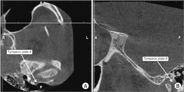
Fig. 1 Left side tympanic plate fracture. A. Axial view. B. Sagittal view.

Fig. 2 Right side tympanic plate fracture. A. Axial view. B. Coronal view. C. Sagittal view.
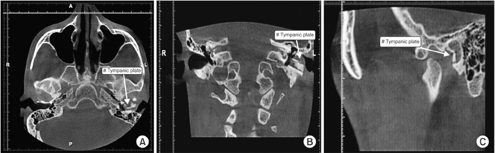
Fig. 3 Left side tympanic plate fracture. A. Axial view. B. Coronal view. C. Sagittal view.

Fig. 4 Right side tympanic plate fracture (TPF). A. Coronal view. B. Sagittal view.
Reference
-
1. Chong VF, Fan YF. Technical report. External auditory canal fracture secondary to mandibular trauma. Clin Radiol. 2000; 55:714–716.2. Gomes MB, Guimarães SM, Filho RG, Neves AC. Traumatic fractures of the tympanic plate: a literature review and case report. Cranio. 2007; 25:134–137.
Article3. Conover GL, Crammond RJ. Tympanic plate fracture from mandibular trauma. J Oral Maxillofac Surg. 1985; 43:292–294.
Article4. Andrade Filho EF, Fadul Junior R, Azevedo RA, Rocha MA, Santos Rd, Toledo SR, et al. Mandibular fractures: analysis of 166 cases. Rev Assoc Med Bras (1992). 2000; 46:272–276.5. Rappaport NH, Scholl PD, Harris JH Jr. Injury to the glenoid fossa. Plast Reconstr Surg. 1986; 77:304–308.
Article6. Copenhaver RH, Dennis MJ, Kloppedal E, Edwards DB, Scheffer RB. Fracture of the glenoid fossa and dislocation of the mandibular condyle into the middle cranial fossa. J Oral Maxillofac Surg. 1985; 43:974–977.
Article7. Pepper L, Zide MF. Mandibular condyle fracture and dislocation into the middle cranial fossa. Int J Oral Surg. 1985; 14:278–283.
Article8. Loh FC, Tan KB, Tan KK. Auditory canal haemorrhage following mandibular condylar fracture. Br J Oral Maxillofac Surg. 1991; 29:12–13.
Article9. Harwood-Nash DC. Fractures of the petrous and tympanic parts of the temporal bone in children: a tomographic study of 35 cases. Am J Roentgenol Radium Ther Nucl Med. 1970; 110:598–607.
Article10. Wood CP, Hunt CH, Bergen DC, Carlson ML, Diehn FE, Schwartz KM, et al. Tympanic plate fractures in temporal bone trauma: prevalence and associated injuries. AJNR Am J Neuroradiol. 2014; 35:186–190.
Article11. Zayas JO, Feliciano YZ, Hadley CR, Gomez AA, Vidal JA. Temporal bone trauma and the role of multidetector CT in the emergency department. Radiographics. 2011; 31:1741–1755.
Article12. Brodie HA, Thompson TC. Management of complications from 820 temporal bone fractures. Am J Otol. 1997; 18:188–197.13. Kim YH, Kim MK, Kang SH. Anterior tympanic plate fracture following extraction of the lower molar. J Korean Assoc Oral Maxillofac Surg. 2016; 42:51–54.
Article14. Antoniades K, Karakasis D, Daggilas A. Posterior dislocation of mandibular condyle into external auditory canal. A case report. Int J Oral Maxillofac Surg. 1992; 21:212–214.
Article15. Psimopoulou M, Antoniades K, Magoudi D, Karakasis D. Tympanic plate fracture following mandibular trauma. Dentomaxillofac Radiol. 1997; 26:344–346.
Article16. Goldberg MH, Aslanian R, Wright J, Marco W. Auditory canal hemorrhage--a sign of mandibular trauma: report of cases. J Oral Surg. 1971; 29:425–427.17. Altay C, Erdoğan N, Batkı O, Eren E, Altay S, Karasu S, et al. Isolated tympanic plate fracture frequency and its relationship to mandibular trauma. Can Assoc Radiol J. 2014; 65:360–365.
Article18. Thor A, Birring E, Leiggener C. Fracture of the tympanic plate with soft tissue extension into the auditory canal resulting from an unfavorable chewing experience. Dent Traumatol. 2010; 26:112–114.
Article19. Dahmani-Causse M, Marx M, Deguine O, Fraysse B, Lepage B, Escudé B. Morphologic examination of the temporal bone by cone beam computed tomography: comparison with multislice helical computed tomography. Eur Ann Otorhinolaryngol Head Neck Dis. 2011; 128:230–235.
Article20. Okeson JP. Management of temporomandibular disorders and occlusion. 7th ed. St. Louis: Elsevier;2012.
- Full Text Links
-
- Actions
-
Cited
- CITED
-
- Close
- Share
-
- Similar articles
-
- Cone beam CT findings of retromolar canals: Report of cases and literature review
- Detection of maxillary second molar with two palatal roots using cone beam computed tomography: a case report
- Three-dimensional imaging modalities in endodontics
- Management of root canal perforation by using cone-beam computed tomography
- Cone beam computed tomography findings of ectopic mandibular third molar in the mandibular condyle: report of a case
