Endovascular Treatment of a Fusiform Aneurysm Involving a Premammillary Artery Originating from the Internal Carotid Artery: A Case Report
- Affiliations
-
- 1Department of Neurosurgery, Inha University School of Medicine and Hospital, Incheon, Korea. dro3@nate.com
- KMID: 2393502
- DOI: http://doi.org/10.7461/jcen.2017.19.3.196
Abstract
- The premammillary artery (PMA) is a branch of the posterior communicating artery (PCoA). While the PMA is known to originate from the PCoA as demonstrated by most anatomical studies, it originates directly from the internal carotid artery in approximately 1% of patients. Cerebral aneurysms associated with the PMA have rarely been reported. We report an extremely rare case of a ruptured PMA aneurysm that was managed using endovascular treatment.
Keyword
Figure
-
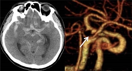
Fig. 1 Computed tomography (CT) of the brain demonstrating subarachnoid hemorrhage (SAH) within the basal cisterns with an intra-parenchymal hematoma observed extending to the left frontal lobe. A CT angiogram demonstrates a fusiform aneurysm involving the communicating segment of the right internal carotid artery.
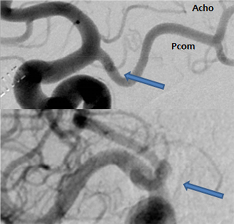
Fig. 2 Digital subtraction angiography (DSA) reveals an aneurysm involving the proximal segment of an undefined parent artery (thick arrows) originating from the internal carotid artery (ICA). The parent artery originates from the ICA just proximal to the origin of the posterior communicating artery (PCoA). This parent artery shows a short course in its distal segment running toward the prepontine cistern in a medial-posterior direction. The artery does not form fenestrations with the PCoA and anterior choroidal artery and follows an independent course.
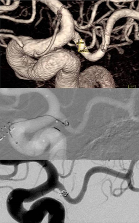
Fig. 3 An Excelsior SL-10 microcatheter (Stryker, Fremont, CA, USA) is navigated into the premammillary artery with its tip positioned in the aneurysm. The aneurysm is subsequently occluded using 2 detachable coils.
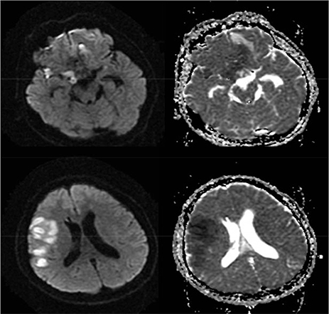
Fig. 4 Diffusion-weighted magnetic resonance imaging (MRI) performed on postoperative day 11 showing high signal intensity within the right hypothalamus and fronto-parietal lobe. A correlative apparent diffusion coefficient (ADC) hypointensity is demonstrated within the same area.
Reference
-
1. Bogousslavsky J, Regli F, Assal G. The syndrome of unilateral tuberothalamic artery territory infarction. Stroke. 1986; May-Jun. 17(3):434–441.
Article2. Endo H, Sato K, Kondo R, Matsumoto Y, Takahashi A, Tominaga T. Tuberothalamic artery infarctions following coil embolization of ruptured posterior communicating artery aneurysms with posterior communicating artery sacrifice. AJNR Am J Neuroradiol. 2012; 03. 33(3):500–506.
Article3. Gabrovsky S, Laleva M, Gabrovsky N. The premammillary artery-a microanatomical study. Acta Neurochir (Wien). 2010; 12. 152(12):2183–2189.4. Gibo H, Lenkey C, Rhoton AL Jr. Microsurgical anatomy of the supraclinoid portion of the internal carotid artery. J Neurosurg. 1981; 10. 55(4):560–574.
Article5. Gibo H, Marinkovic S, Brigante L. The microsurgical anatomy of the premammillary artery. J Clin Neurosci. 2001; 05. 8(3):256–260.6. Kim DS, Yoo DS, Huh PW, Cho KS, Kang JK. Anterior thalamoperforating artery aneurysm associated with internal carotid artery occlusion: case report. Neurosurgery. 1999; 10. 45(4):911–913.
Article7. Kownacki JD, Remonda L, Godoy N, Krauss J. Subependymal thalamic haemorrhage due to a thalamoperforating artery aneurysm. J Neurol Neurosurg Psychiatry. 1998; 11. 65(5):669. 678.
Article8. Kumar AJ, Zinreich SJ, Preziosi TJ. Rupture of an anterior thalamoperforating artery aneurysm: cause of basal ganglia hemorrhage. AJNR Am J Neuroradiol. 1982; Sep-Oct. 3(5):581–582.9. Kim SH, Yeo DK, Shum JJ, Yoon SM, Chang JC, Bae HG. Morphometric Study of the Anterior thalamoperforating Arteries. J Korean Neurosurg Soc. 2015; 05. 57(5):350–358.
Article10. Lee JI, Choi CH, Ko JK, Lee TH. Glue embolization of ruptured anterior thalamoperforating artery aneurysm in patient with both internal carotid arteries occlusion. J Korean Neurosurg Soc. 2011; 05. 49(5):287–289.
Article11. Lisovoski F, Koskas P, Dubard T, Dessarts I, Dehen H, Cambier J. Left tuberothalamic artery territory infarction: neuropsychological and MRI features. Eur Neurol. 1993; 33(2):181–184.
Article12. Pedroza A, Dujovny M, Cabezudo-Artero J, Umansky F, Berman SK, Diaz FG, et al. Microanatomy of the premammillary artery. Acta Neurochir (Wien). 1987; 86(1-2):50–55.13. Saeki N, Rhoton AL Jr. Microsurgical anatomy of the upper basilar artery and the posterior circle of Willis. J Neurosurg. 1977; 05. 46(5):563–578.
Article
- Full Text Links
-
- Actions
-
Cited
- CITED
-
- Close
- Share
-
- Similar articles
-
- Stent-Assisted Coil Trapping in a Manual Internal Carotid Artery Compression Test for the Treatment of a Fusiform Dissecting Aneurysm
- Supraclinoid Internal Carotid Artery Fenestration Harboring an Unruptured Aneurysm and Another Remote Ruptured Aneurysm: Case Report and Review of the Literature
- Serpentine Cavernous Aneurysm Presented with Visual Symptoms Improved by Endovascular Coil Trapping
- Dual Stent-Assisted Coil Embolization for Fusiform Aneurysm Arising From Persistent Trigeminal Artery
- Fusiform “True” Posterior Communicating Artery Aneurysm with Basilar Artery Occlusion: A Case Report
