Serpentine Cavernous Aneurysm Presented with Visual Symptoms Improved by Endovascular Coil Trapping
- Affiliations
-
- 1Department of Neurosurgery, Soonchunhyang University Bucheon Hospital, Bucheon, Korea. bumtkim@schmc.ac.kr
- 2Department of Neurosurgery, Preah Ket Mealea Hospital, Phnom Pehn, Cambodia.
- KMID: 2367330
- DOI: http://doi.org/10.7461/jcen.2016.18.4.379
Abstract
- This report describes a case of a serpentine fusiform aneurysm of the internal carotid artery in a patient who presented with visual disturbances. The serpentine aneurysm was treated successfully by coil trapping and occlusion of the parent artery, accompanied by balloon dilation. Nine months post-operatively, the patient's visual acuity had improved considerably.
Figure
-
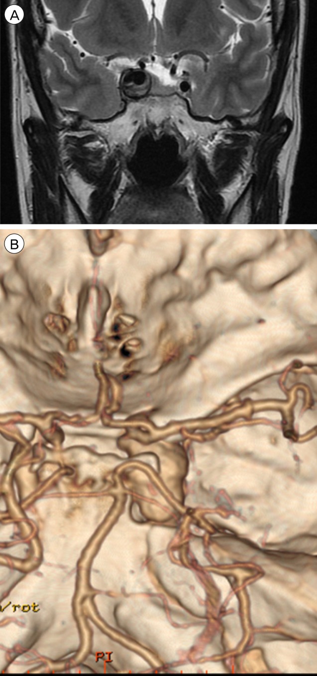
Fig. 1 (A) MRI shows a large thrombosed aneurysm 19 × 12 mm in size, well marginated, and oval-shaped, with a heterogeneous signal void in the right parasellar region, probably in the cavernous sinus. (B) CTA shows a fusiform dilatation of the right cavernous ICA, suggestive of a fusiform aneurysm approximately 30 × 20 mm in size. MRI = magnetic resonance imaging; CTA = computed tomography angiography; ICA = internal carotid artery.
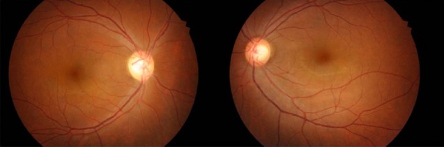
Fig. 2 Fundus photography shows right optic nerve atrophy, with a pale neural rim on the right optic disc.
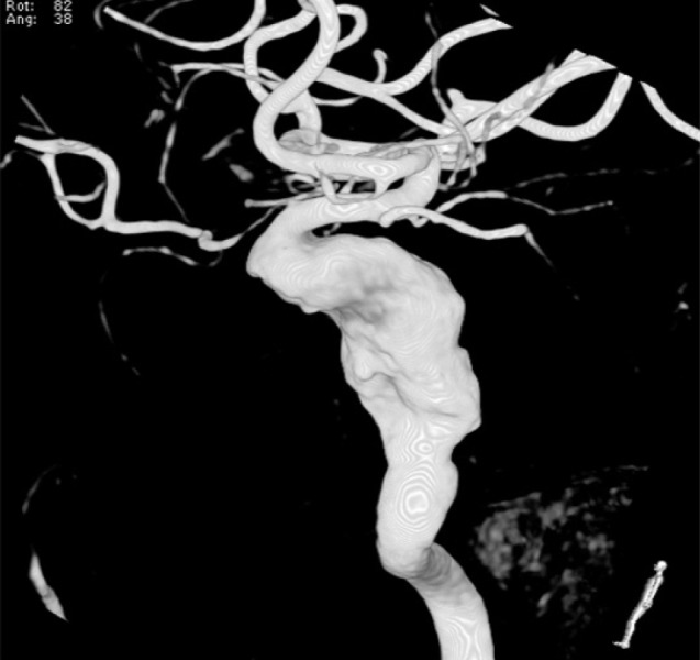
Fig. 3 3D angiography shows a large serpentine aneurysm of the right ICA 11.72 × 22.49 mm in size. 3D = three-dimensional; ICA = internal carotid artery.

Fig. 4 (A, B) Matas test performed on the right ICA using balloon dilation of the parent artery followed by observation of the patient for 20 min. (C) SPECT shows the well-maintained vascular reserve. (D, E-1, E-2) Collateral flow from the left ICA & vertebrobasilar system was well visualized on preoperative cerebral angiography. ICA = internal carotid artery; SPECT = single-photon emission computed tomography.

Fig. 5 (A-D) Coil embolization with occlusion of the parent artery using balloon dilation was performed using detachable and pushable coils.
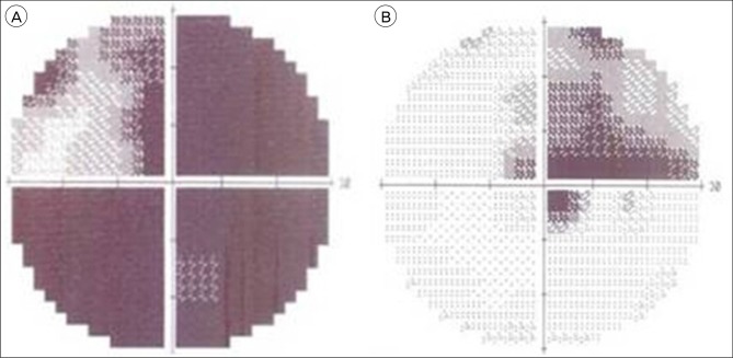
Fig. 6 (A, B) The visual field defect improved 9 months after coil embolization of the aneurysm.
Cited by 1 articles
-
Abducens Nerve Palsy Caused by the Ophthalmic Segment of an Internal Carotid Artery Aneurysm
Inhye Kim, Jong Hoon Kim, Won Jae Kim
J Korean Ophthalmol Soc. 2018;59(4):388-392. doi: 10.3341/jkos.2018.59.4.388.
Reference
-
1. Aletich VA, Debrun GM, Monsein LH, Nauta HJ, Spetzler RF. Giant serpentine aneurysms: a review and presentation of five cases. AJNR Am J Neuroradiol. 1995; 5. 16(5):1061–1072. PMID: 7639128.2. Drake CG, Peerless SJ. Giant fusiform intracranial aneurysms: review of 120 patients treated surgically from 1965 to 1992. J Neurosurg. 1997; 8. 87(2):141–162. PMID: 9254076.
Article3. Greene KA, Anson JA, Spetzler RF. Giant serpentine middle cerebral artery aneurysm treated by extracranial-intracranial bypass. Case report. J Neurosurg. 1993; 6. 78(6):974–978. PMID: 8487082.4. Haddad GF, Haddad FS. Cerebral giant serpentine aneurysm: case report and review of the literature. Neurosurgery. 1988; 7. 23(1):92–97. PMID: 3050586.
Article5. Halbach VV, Higashida RT, Dowd CF, Barnwell SL, Fraser KW, Smith TP, et al. The efficacy of endosaccular aneurysm occlusion in alleviating neurological deficits produced by mass effect. J Neurosurg. 1994; 4. 80(4):659–666. PMID: 8151344.
Article6. Higashida RT, Halbach VV, Dowd C, Barnwell SL, Dormandy B, Bell J, et al. Endovascular detachable balloon embolization therapy of cavernous carotid artery aneurysms: results in 87 cases. J Neurosurg. 1990; 6. 72(6):857–863. PMID: 2338569.
Article7. Larson JJ, Tew JM Jr, Tomsick TA, van Loveren HR. Treatment of aneurysms of the internal carotid artery by intravascular balloon occlusion: long-term follow-up of 58 patients. Neurosurgery. 1995; 1. 36(1):26–30. discussion 30. PMID: 7708164.
Article8. Lee KC, Joo JY, Lee KS, Shin YS. Recanalization of completely thrombosed giant aneurysm: case report. Surg Neurol. 1999; 1. 51(1):94–98. PMID: 9952130.
Article9. Lukin RR, Chambers AA, McLaurin R, Tew J Jr. Thrombosed giant middle cerebral aneurysms. Neuroradiology. 1975; 12. 10(3):125–129. PMID: 1207884.
Article10. Malisch TW, Guglielmi G, Vinuela F, Duckwiler G, Gobin YP, Martin NA, et al. Unruptured aneurysms presenting with mass effect symptoms: response to endosaccular treatment with Guglielmi detachable coils. Part I. Symptoms of cranial nerve dysfunction. J Neurosurg. 1998; 12. 89(6):956–961. PMID: 9833822.11. Mawad ME, Klucznik RP. Giant serpentine aneurysms: radiographic features and endovascular treatment. AJNR Am J Neuroradiol. 1995; 5. 16(5):1053–1060. PMID: 7639127.12. Mizutani T, Aruga T, Kirino T, Miki Y, Saito I, Tsuchida T. Recurrent subarachnoid hemorrhage from untreated ruptured vertebrobasilar dissecting aneurysms. Neurosurgery. 1995; 5. 36(5):905–911. discussion 912-3. PMID: 7791980.
Article13. Patel DV, Sherman IC, Hemmati M, Ferguson RJ. Giant serpentine intracranial aneurysm. Surg Neurol. 1981; 12. 16(6):402–407. PMID: 7330759.
Article14. Segal HD, McLaurin RL. Giant serpentine aneurysm. Report of two cases. J Neurosurg. 1977; 1. 46(1):115–120. PMID: 830809.15. Strother CM, Eldevik P, Kikuchi Y, Graves V, Partington C, Merlis A. Thrombus formation and structure and the evolution of mass effect in intracranial aneurysms treated by balloon embolization: emphasis on MR findings. AJNR Am J Neuroradiol. 1989; Jul-Aug. 10(4):787–796. PMID: 2505506.16. Tomasello F, Albanese V, Cioffi FA. Giant serpentine aneurysms: a separate entity. Surg Neurol. 1979; 11. 12(5):429–432. PMID: 515943.
- Full Text Links
-
- Actions
-
Cited
- CITED
-
- Close
- Share
-
- Similar articles
-
- Endovascular occlusion of giant serpentine aneurysm: A case report and literature review
- Endovascular Coil Trapping of a Ruptured Dissecting Aneurysm of the Vertebral Artery Using Detachable Coils and Micro-Tornado(R) Coils
- Giant Serpentine Aneurysm of the Posterior Cerebral Artery: Case Report
- Stent-Assisted Coil Trapping in a Manual Internal Carotid Artery Compression Test for the Treatment of a Fusiform Dissecting Aneurysm
- Recurrent Carotid Cavernous Fistula Originating from a Giant Cerebral Aneurysm after Placement of a Covered Stent
