Progression of Right Ventricular Systolic Dysfunction Detected by Myocardial Deformation Imaging in Asymptomatic Preterm Children
- Affiliations
-
- 1Department of Pediatrics, CHA Bundang Medical Center, CHA University School of Medicine, Seongnam, Korea. kittysooni@chamc.co.kr
- 2Department of Diagnostic Laboratory Medicine, CHA Bundang Medical Center, CHA University School of Medicine, Seongnam, Korea.
- KMID: 2392255
- DOI: http://doi.org/10.4250/jcu.2017.25.3.98
Abstract
- BACKGROUND
To detect progression of right ventricular (RV) systolic dysfunction (RVSD) in asymptomatic preterm children from infancy to 24-month corrected age, using velocity vector imaging (VVI).
METHODS
Retrospective study comparing sequential RV longitudinal peak systolic strain (LPSS) from 24 children born at < 33 weeks of gestational age and 10 term infants recruited as controls, obtained at a mean of 4-month (first exam) and 24-month corrected age (second exam).
RESULTS
In 7/24 (29.2%) of preterm children, RV LPSS of < 16%, defined as RVSD, was detected at the second exam; 5/7 of these children had RV LPSS > 16% at the first exam, and only 2/7 of these children had a history of moderate or severe bronchopulmonary dysplasia.
CONCLUSION
In asymptomatic preterm children, routine echocardiographic screening using VVI could detect RVSD which could progress from 4-24 month corrected age.
MeSH Terms
Figure
-

Fig. 1 Example of right ventricular longitudinal peak systolic strain (RV LPSS) identified as the highest point of the average strain curve derived from six segments observed using velocity vector imaging in a preterm child.

Fig. 2 Change over time in right ventricular longitudinal peak systolic strain (RV LPSS) in preterm children and controls from a mean 4- to 24-month corrected age. LPSS1: RV LPSS at a mean 4 month corrected age, LPSS2: RV LPSS at a mean 24 month corrected age.
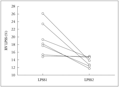
Fig. 3 Change over time in right ventricular longitudinal peak systolic strain (RV LPSS) in individual preterm children with right ventricular systolic dysfunction at a mean 24 month corrected age. LPSS1: RV LPSS at a mean 4 month corrected age, LPSS2: RV LPSS at a mean 24 month corrected age.
Reference
-
1. Breatnach CR, Levy PT, James AT, Franklin O, El-Khuffash A. Novel echocardiography methods in the functional assessment of the newborn heart. Neonatology. 2016; 110:248–260.2. Bhat R, Salas AA, Foster C, Carlo WA, Ambalavanan N. Prospective analysis of pulmonary hypertension in extremely low birth weight infants. Pediatrics. 2012; 129:e682–e689.3. Mourani PM, Sontag MK, Younoszai A, Miller JI, Kinsella JP, Baker CD, Poindexter BB, Ingram DA, Abman SH. Early pulmonary vascular disease in preterm infants at risk for bronchopulmonary dysplasia. Am J Respir Crit Care Med. 2015; 191:87–95.4. Levy PT, El-Khuffash A, Patel MD, Breatnach CR, James AT, Sanchez AA, Abuchabe C, Rogal SR, Holland MR, McNamara PJ, Jain A, Franklin O, Mertens L, Hamvas A, Singh GK. Maturational patterns of systolic ventricular deformation mechanics by two-dimensional speckletracking echocardiography in preterm infants over the first year of age. J Am Soc Echocardiogr. 2017; 30:685–698.5. Farrow KN, Steinhorn RH. Pulmonary hypertension in premature infants. Sharpening the tools of detection. Am J Respir Crit Care Med. 2015; 191:12–14.6. Mourani PM, Sontag MK, Younoszai A, Ivy DD, Abman SH. Clinical utility of echocardiography for the diagnosis and management of pulmonary vascular disease in young children with chronic lung disease. Pediatrics. 2008; 121:317–325.7. Petitjean C, Rougon N, Cluzel P. Assessment of myocardial function: a review of quantification methods and results using tagged MRI. J Cardiovasc Magn Reson. 2005; 7:501–516.8. Levy PT, Holland MR, Sekarski TJ, Hamvas A, Singh GK. Feasibility and reproducibility of systolic right ventricular strain measurement by speckle-tracking echocardiography in premature infants. J Am Soc Echocardiogr. 2013; 26:1201–1213.9. Haque U, Stiver C, Rivera BK, Richards B, Ma N, Cua CL, Smith CV, Backes CH. Right ventricular performance using myocardial deformation imaging in infants with bronchopulmonary dysplasia. J Perinatol. 2017; 37:81–87.10. Nestaas E, Støylen A, Brunvand L, Fugelseth D. Longitudinal strain and strain rate by tissue Doppler are more sensitive indices than fractional shortening for assessing the reduced myocardial function in asphyxiated neonates. Cardiol Young. 2011; 21:1–7.11. Pirat B, Khoury DS, Hartley CJ, Tiller L, Rao L, Schulz DG, Nagueh SF, Zoghbi WA. A novel feature-tracking echocardiographic method for the quantitation of regional myocardial function: validation in an animal model of ischemia-reperfusion. J Am Coll Cardiol. 2008; 51:651–659.12. Engle WA. American Academy of Pediatrics Committee on Fetus and Newborn. Age terminology during the perinatal period. Pediatrics. 2004; 114:1362–1364.13. Jobe AH, Bancalari E. Bronchopulmonary dysplasia. Am J Respir Crit Care Med. 2001; 163:1723–1729.14. Lai WW, Geva T, Shirali GS, Frommelt PC, Humes RA, Brook MM, Pignatelli RH, Rychik J. Task Force of the Pediatric Council of the American Society of Echocardiography. Pediatric Council of the American Society of Echocardiography. Guidelines and standards for performance of a pediatric echocardiogram: a report from the Task Force of the Pediatric Council of the American Society of Echocardiography. J Am Soc Echocardiogr. 2006; 19:1413–1430.15. Lang RM, Bierig M, Devereux RB, Flachskampf FA, Foster E, Pellikka PA, Picard MH, Roman MJ, Seward J, Shanewise JS, Solomon SD, Spencer KT, Sutton MS, Stewart WJ. Chamber Quantification Writing Group. American Society of Echocardiography's Guidelines and Standards Committee. European Association of Echocardiography. Recommendations for chamber quantification: a report from the American Society of Echocardiography’s Guidelines and Standards Committee and the Chamber Quantification Writing Group, developed in conjunction with the European Association of Echocardiography, a branch of the European Society of Cardiology. J Am Soc Echocardiogr. 2005; 18:1440–1463.16. Sehgal A, Malikiwi A, Paul E, Tan K, Menahem S. Right ventricular function in infants with bronchopulmonary dysplasia: association with respiratory sequelae. Neonatology. 2016; 109:289–296.17. Lopez L, Colan SD, Frommelt PC, Ensing GJ, Kendall K, Younoszai AK, Lai WW, Geva T. Recommendations for quantification methods during the performance of a pediatric echocardiogram: a report from the Pediatric Measurements Writing Group of the American Society of Echocardiography Pediatric and Congenital Heart Disease Council. J Am Soc Echocardiogr. 2010; 23:465–495.18. Lang RM, Badano LP, Mor-Avi V, Afilalo J, Armstrong A, Ernande L, Flachskampf FA, Foster E, Goldstein SA, Kuznetsova T, Lancellotti P, Muraru D, Picard MH, Rietzschel ER, Rudski L, Spencer KT, Tsang W, Voigt JU. Recommendations for cardiac chamber quantification by echocardiography in adults: an update from the American Society of Echocardiography and the European Association of Cardiovascular Imaging. J Am Soc Echocardiogr. 2015; 28:1–39.19. Levy PT, Patel MD, Groh G, Choudhry S, Murphy J, Holland MR, Hamvas A, Grady MR, Singh GK. Pulmonary artery acceleration time provides a reliable estimate of invasive pulmonary hemodynamics in children. J Am Soc Echocardiogr. 2016; 29:1056–1065.20. Voigt JU, Pedrizzetti G, Lysyansky P, Marwick TH, Houle H, Baumann R, Pedri S, Ito Y, Abe Y, Metz S, Song JH, Hamilton J, Sengupta PP, Kolias TJ, d'Hooge J, Aurigemma GP, Thomas JD, Badano LP. Definitions for a common standard for 2D speckle tracking echocardiography: consensus document of the EACVI/ASE/Industry Task Force to standardize deformation imaging. J Am Soc Echocardiogr. 2015; 28:183–193.21. Kutty S, Padiyath A, Li L, Peng Q, Rangamani S, Schuster A, Danford DA. Functional maturation of left and right atrial systolic and diastolic performance in infants, children, and adolescents. J Am Soc Echocardiogr. 2013; 26:398–409.22. Poets CF, Stebbens VA, Richard D, Southall DP. Prolonged episodes of hypoxemia in preterm infants undetectable by cardiorespiratory monitors. Pediatrics. 1995; 95:860–863.23. Schubert U, Müller M, Abdul-Khaliq H, Norman M. Preterm birth is associated with altered myocardial function in infancy. J Am Soc Echocardiogr. 2016; 29:670–678.24. Sanchez AA, Levy PT, Sekarski TJ, Hamvas A, Holland MR, Singh GK. Effects of frame rate on two-dimensional speckle tracking-derived measurements of myocardial deformation in premature infants. Echocardiography. 2015; 32:839–847.25. Levy PT, Sanchez Mejia AA, Machefsky A, Fowler S, Holland MR, Singh GK. Normal ranges of right ventricular systolic and diastolic strain measures in children: a systematic review and meta-analysis. J Am Soc Echocardiogr. 2014; 27:549–560.
- Full Text Links
-
- Actions
-
Cited
- CITED
-
- Close
- Share
-
- Similar articles
-
- Sequential Changes in Left Ventricular Systolic Myocardial Deformation Mechanics in Children with Recurrent Kawasaki Disease
- Principles and Clinical Applications of Feature-Tracking Cardiac Magnetic Resonance Imaging: A Literature Review
- Functional Assessment for Congenital Heart Disease
- Strain Analysis of the Right Ventricle Using Two-dimensional Echocardiography
- The Usefulness of Doppler Tissue Image in Evaluation of Left Ventricular Systolic and Diastolic Dysfunction
