Effects of bodily retraction of mandibular incisors versus mandibular setback surgery on pharyngeal airway space: A comparative study
- Affiliations
-
- 1Department of Orthodontics, College of Dentistry, Yonsei University, Seoul, Korea. orthojn@yuhs.ac
- 2Institute of Craniofacial Deformity, College of Dentistry, Yonsei University, Seoul, Korea.
- KMID: 2392181
- DOI: http://doi.org/10.4041/kjod.2017.47.6.344
Abstract
OBJECTIVE
The purpose of this study was to compare the changes induced in the pharyngeal airway space by orthodontic treatment with bodily retraction of the mandibular incisors and mandibular setback surgery without extraction.
METHODS
This retrospective study included 63 adult patients (32 men and 31 women). Thirty-three patients who had been treated via four-bicuspid extraction and bodily retraction of the mandibular incisors (incisor retraction, IR group) were compared with 30 patients who had been treated via mandibular setback surgery (MS group) without extraction. Lateral cephalograms were acquired and analyzed before (T1) and after treatment (T2).
RESULTS
The superior pharyngeal airway space did not change significantly in either group during treatment. The middle pharyngeal airway space decreased by 1.15 ± 1.17 mm and 1.25 ± 1.35 mm after treatment in the IR and MS groups, respectively, and the decrease was comparable between the two groups. In the MS group, the inferior pharyngeal airway space (E-IPW) decreased by 0.88 ± 1.67 mm after treatment (p < 0.01). The E-IPW was larger in the MS group than in IR group at T1, but it did not differ significantly between the two groups at T2. No significant correlation was observed between changes in the pharyngeal airway space and the skeletal and dental variables in each group.
CONCLUSIONS
The middle pharyngeal airway space decreased because of the posterior displacement of the mandibular incisors and/or the mandibular body. The E-IPW decreased only in the MS group because of the posterior displacement of only the mandibular body.
MeSH Terms
Figure
-
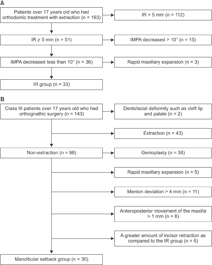
Figure 1 Flow diagram illustrating sample selection. IR, Incisor retraction; IMPA, incisor mandibular plane angle.
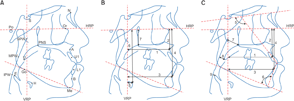
Figure 2 A, Cephalometric landmarks and planes. S, sella; N,nasion; A, point A; B, point B; Po, porion; Or, orbitale; PNS, posterior nasal spine; Go, gonion; Me, menton; H, the most anterosuperior point of the hyoid bone; U, tip of the uvula; E, tip of the epiglottis; U1, center of resistance of U1; L1, center of resistance of L1; HRP, horizontal reference plane–the Frankfort horizontal plane; VRP, vertical reference plane—passes through the sella, perpendicular to the HRP; SPW, superior pharyngeal wall, point of intersection of the posterior pharyngeal wall and perpendicular line drawn from the PNS; MPW, middle pharyngeal wall, point of intersection of the posterior pharyngeal wall and perpendicular line drawn from the U; IPW, inferior pharyngeal wall, point of intersection of the posterior pharyngeal wall and perpendicular line drawn from the E. B, Skeletal measurements. 1, A-VRP, perpendicular distance from the VRP to point A (mm); 2, A-HRP, perpendicular distance from the HRP to point A (mm); 3, B-VRP, perpendicular distance from the VRP to point B (mm); 4, B-HRP, perpendicular distance from the HRP to point B (mm); 5, H-VRP, perpendicular distance from the VRP to point H (mm); 6, H-HRP, perpendicular distance from the HRP to point H (mm); 7, PNS-HRP, perpendicular distance from the HRP to the PNS (mm). C, Dental and pharyngeal airway space measurements. 1, U1-VRP, perpendicular distance from the VRP to U1 (mm); 2, U1-HRP, perpendicular distance from the HRP to U1 (mm); 3, L1-VRP, perpendicular distance from the VRP to L1 (mm); 4, L1-HRP, perpendicular distance from the HRP to L1 (mm); 5, U1-SN (o); 6, IMPA (o); 7, PNS-SPW, superior pharyngeal airway (mm); 8, U-MPW, middle pharyngeal airway (mm); 9, E-IPW, inferior pharyngeal airway (mm).
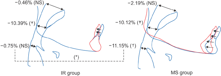
Figure 3 Changes in pharyngeal airway space during treatment. Blue line, before treatment; red line, after treatment. NS, Not significant; IR, incisor retraction; MS, mandibular setback surgery. *p < 0.05; †p < 0.01; ‡p < 0.001.
Cited by 1 articles
-
Effect of extraction treatment on upper airway dimensions in patients with bimaxillary skeletal protrusion relative to their vertical skeletal pattern
Ha-Nul Cho, Hyun Joo Yoon, Jae Hyun Park, Young-Guk Park, Su-Jung Kim
Korean J Orthod. 2021;51(3):166-178. doi: 10.4041/kjod.2021.51.3.166.
Reference
-
1. Hoekema A, Hovinga B, Stegenga B, De Bont LG. Craniofacial morphology and obstructive sleep apnoea: a cephalometric analysis. J Oral Rehabil. 2003; 30:690–696.
Article2. Alessandri-Bonetti G, Ippolito DR, Bartolucci ML, D'Antò V, Incerti-Parenti S. Cephalometric predictors of treatment outcome with mandibular advancement devices in adult patients with obstructive sleep apnea: a systematic review. Korean J Orthod. 2015; 45:308–321.
Article3. Tselnik M, Pogrel MA. Assessment of the pharyngeal airway space after mandibular setback surgery. J Oral Maxillofac Surg. 2000; 58:282–285. discussion 285-7.
Article4. Kawakami M, Yamamoto K, Fujimoto M, Ohgi K, Inoue M, Kirita T. Changes in tongue and hyoid positions, and posterior airway space following mandibular setback surgery. J Craniomaxillofac Surg. 2005; 33:107–110.
Article5. Hwang S, Chung CJ, Choi YJ, Huh JK, Kim KH. Changes of hyoid, tongue and pharyngeal airway after mandibular setback surgery by intraoral vertical ramus osteotomy. Angle Orthod. 2010; 80:302–308.
Article6. Gokce SM, Gorgulu S, Gokce HS, Bengi O, Sabuncuoglu F, Ozgen F, et al. Changes in posterior airway space, pulmonary function and sleep quality, following bimaxillary orthognathic surgery. Int J Oral Maxillofac Surg. 2012; 41:820–829.
Article7. Mattos CT, Vilani GN, Sant'Anna EF, Ruellas AC, Maia LC. Effects of orthognathic surgery on oropharyngeal airway: a meta-analysis. Int J Oral Maxillofac Surg. 2011; 40:1347–1356.
Article8. Park HS, Yoon DY, Park CS, Jeoung SH. Treatment effects and anchorage potential of sliding mechanics with titanium screws compared with the Tweed-Merrifield technique. Am J Orthod Dentofacial Orthop. 2008; 133:593–600.
Article9. Liu YH, Ding WH, Liu J, Li Q. Comparison of the differences in cephalometric parameters after active orthodontic treatment applying mini-screw implants or transpalatal arches in adult patients with bialveolar dental protrusion. J Oral Rehabil. 2009; 36:687–695.
Article10. Germec-Cakan D, Taner T, Akan S. Uvulo-glossopharyngeal dimensions in non-extraction, extraction with minimum anchorage, and extraction with maximum anchorage. Eur J Orthod. 2011; 33:515–520.
Article11. Chen Y, Hong L, Wang CL, Zhang SJ, Cao C, Wei F, et al. Effect of large incisor retraction on upper airway morphology in adult bimaxillary protrusion patients. Angle Orthod. 2012; 82:964–970.
Article12. Wang Q, Jia P, Anderson NK, Wang L, Lin J. Changes of pharyngeal airway size and hyoid bone position following orthodontic treatment of Class I bimaxillary protrusion. Angle Orthod. 2012; 82:115–121.
Article13. Burstone CJ, Pryputniewicz RJ. Holographic determination of centers of rotation produced by orthodontic forces. Am J Orthod. 1980; 77:396–409.
Article14. Koo YJ, Choi SH, Keum BT, Yu HS, Hwang CJ, Melsen B, et al. Maxillomandibular arch width differences at estimated centers of resistance: comparison between normal occlusion and skeletal Class III malocclusion. Korean J Orthod. 2017; 47:167–175.
Article15. Chen F, Terada K, Hua Y, Saito I. Effects of bimaxillary surgery and mandibular setback surgery on pharyngeal airway measurements in patients with Class III skeletal deformities. Am J Orthod Dentofacial Orthop. 2007; 131:372–377.
Article16. Degerliyurt K, Ueki K, Hashiba Y, Marukawa K, Nakagawa K, Yamamoto E. A comparative CT evaluation of pharyngeal airway changes in Class III patients receiving bimaxillary surgery or mandibular setback surgery. Oral Surg Oral Med Oral Pathol Oral Radiol Endod. 2008; 105:495–502.
Article17. Jakobsone G, Stenvik A, Espeland L. The effect of maxillary advancement and impaction on the upper airway after bimaxillary surgery to correct Class III malocclusion. Am J Orthod Dentofacial Orthop. 2011; 139:4 Suppl. e369–e376.
Article18. Alves PV, Zhao L, O'Gara M, Patel PK, Bolognese AM. Three-dimensional cephalometric study of upper airway space in skeletal Class II and III healthy patients. J Craniofac Surg. 2008; 19:1497–1507.
Article19. de Freitas MR, Alcazar NM, Janson G, de Freitas KM, Henriques JF. Upper and lower pharyngeal airways in subjects with Class I and Class II malocclusions and different growth patterns. Am J Orthod Dentofacial Orthop. 2006; 130:742–745.
Article20. Martin O, Muelas L, Viñas MJ. Comparative study of nasopharyngeal soft-tissue characteristics in patients with Class III malocclusion. Am J Orthod Dentofacial Orthop. 2011; 139:242–251.
Article21. Hong JS, Oh KM, Kim BR, Kim YJ, Park YH. Three-dimensional analysis of pharyngeal airway volume in adults with anterior position of the mandible. Am J Orthod Dentofacial Orthop. 2011; 140:e161–e169.
Article22. Zheng ZH, Yamaguchi T, Kurihara A, Li HF, Maki K. Three-dimensional evaluation of upper airway in patients with different anteroposterior skeletal patterns. Orthod Craniofac Res. 2014; 17:38–48.
Article23. Yao CC, Lai EH, Chang JZ, Chen I, Chen YJ. Comparison of treatment outcomes between skeletal anchorage and extraoral anchorage in adults with maxillary dentoalveolar protrusion. Am J Orthod Dentofacial Orthop. 2008; 134:615–624.
Article24. Al-Sibaie S, Hajeer MY. Assessment of changes following en-masse retraction with mini-implants anchorage compared to two-step retraction with conventional anchorage in patients with Class II division 1 malocclusion: a randomized controlled trial. Eur J Orthod. 2014; 36:275–283.
Article25. Hu Z, Yin X, Liao J, Zhou C, Yang Z, Zou S. The effect of teeth extraction for orthodontic treatment on the upper airway: a systematic review. Sleep Breath. 2015; 19:441–451.
Article26. Gu G, Gu G, Nagata J, Suto M, Anraku Y, Nakamura K, et al. Hyoid position, pharyngeal airway and head posture in relation to relapse after the mandibular setback in skeletal Class III. Clin Orthod Res. 2000; 3:67–77.
Article27. Güven O, Saraçoğlu U. Changes in pharyngeal airway space and hyoid bone positions after body ostectomies and sagittal split ramus osteotomies. J Craniofac Surg. 2005; 16:23–30.
Article28. Athanasiou AE, Toutountzakis N, Mavreas D, Ritzau M, Wenzel A. Alterations of hyoid bone position and pharyngeal depth and their relationship after surgical correction of mandibular prognathism. Am J Orthod Dentofacial Orthop. 1991; 100:259–265.
Article29. Malkoc S, Usumez S, Nur M, Donaghy CE. Reproducibility of airway dimensions and tongue and hyoid positions on lateral cephalograms. Am J Orthod Dentofacial Orthop. 2005; 128:513–516.
Article
- Full Text Links
-
- Actions
-
Cited
- CITED
-
- Close
- Share
-
- Similar articles
-
- A Cephalometric Study on Changes in Pharyngeal Airway Space, Tongue and Hyoid Bone Positions Following the Surgical Correction of Mandibular Prognathism
- The effects of mandibular setback osteotomy on the oropharyngeal airway space in mandibular prognathic patients
- Changes of Pharyngeal Airway Space after Mandibular Setback Surgery in Computed Tomography Images
- Why most patients do not exhibit obstructive sleep apnea after mandibular setback surgery?
- Changes in the hyoid bone, tongue, and oropharyngeal airway space after mandibular setback surgery evaluated by cone-beam computed tomography
