Change of the Halo Sign and the Grafted Bone according to the Usage of Teriparatide for the Correction Loss due to Screw Loosening after Corrective Osteotomy
- Affiliations
-
- 1Department of Orthopaedic Surgery, Guri Hospital, Hanyang University College of Medicine, Guri, Korea. hyparkys@hanyang.ac.kr
Abstract
- Among osteoporosis medications, Teriparatide is an agent that promotes bone formation and it seems to have an effect, due to an anabolic mechanism, in the early postoperative period after osteosynthesis or joint replacement. But to the best of our knowledge, the effect of teriparatide on pedicle screw loosening has not been previously reported. We report there on a case of pedicle screw loosening after corrective osteotomy in a patient with ankylosing spondylitis with osteoporosis, which was not improved by teriparatide, and we review the related literature.
MeSH Terms
Figure
-
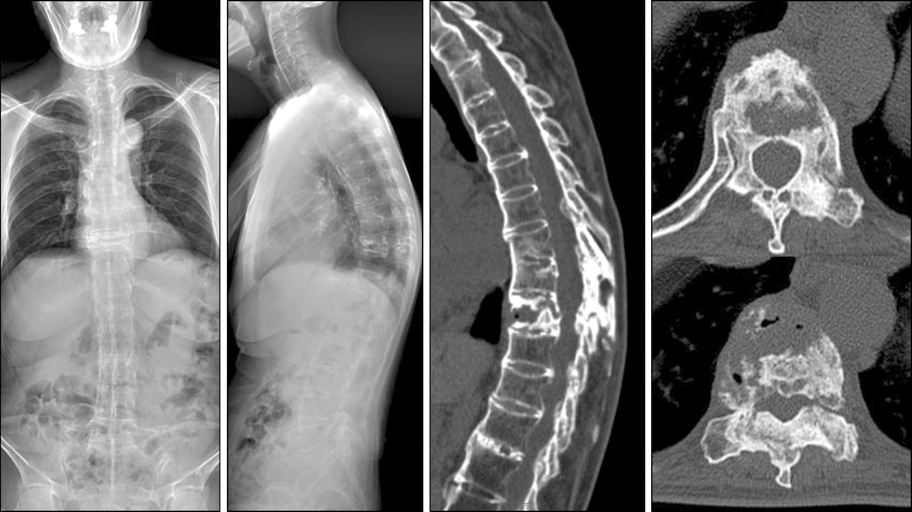
Figure 1. The 60 year-old female had a fixed kyphotic deformity with Andersson lesion due to ankylosing spondylitis and osteoporosis (T-score: −2.6). On the radiologic evaluation, the thoracic kyphosis (TK) was 65 o, the lumbar lordosis (LL) was 12 o and the distance between the C7 plumb line and S1 (C7PL) was 59 mm.
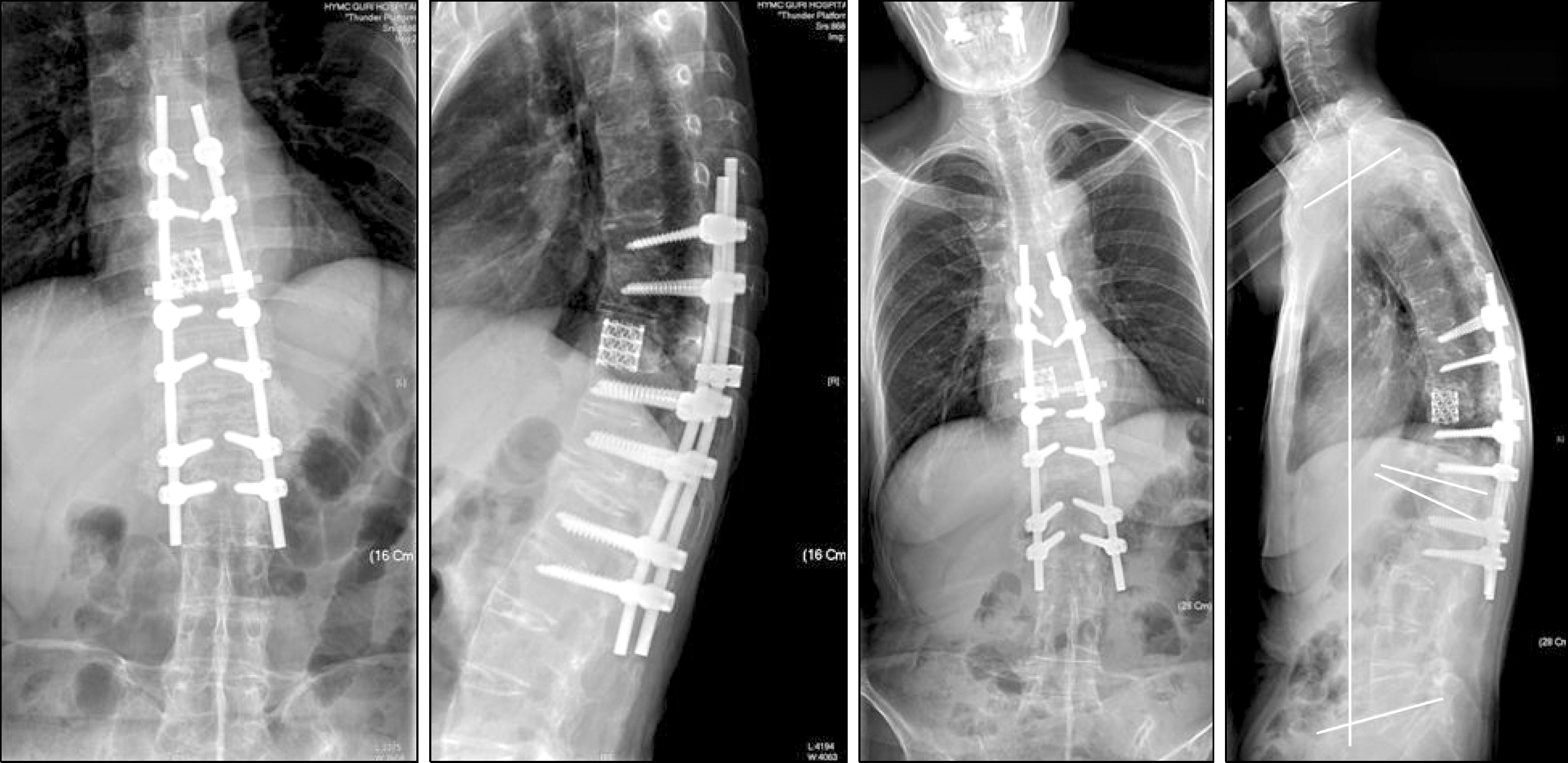
Figure 2. In the first operation, pedicle subtraction osteotomy at T12 and Smith-Petersen osteotomy at T9-10 were done. In the second operation, anterior corpec-tomy of T9 and anterior interbody fusion with Mesh were done in order to treat the Andersson lesion. The TK was 60 o, the LL was −44 o and the C7PL was 33 mm.
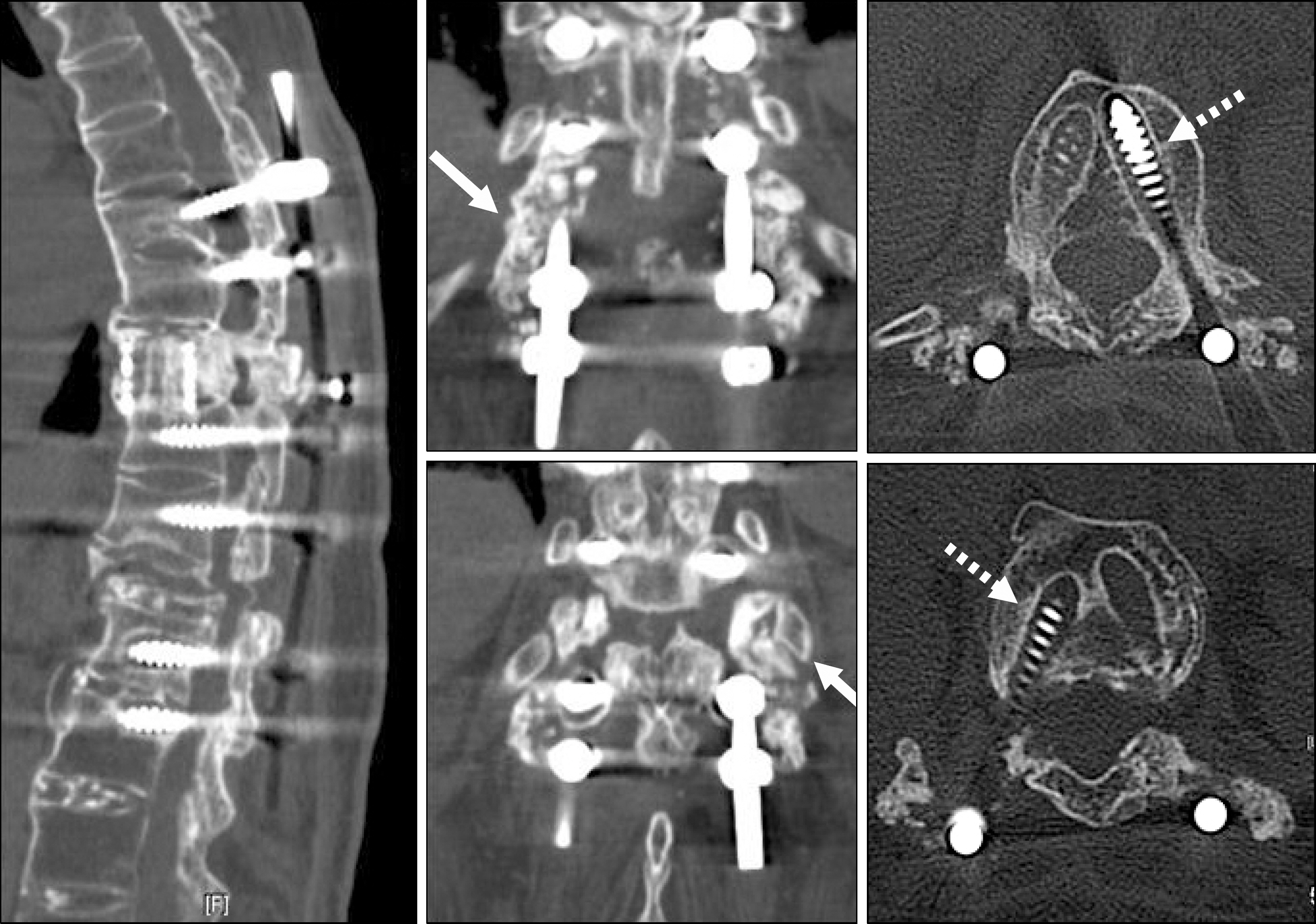
Figure 3. The loss of correction with loosening of screws at L1 and L2 (dot arrows) was seen on the computed tomography. We used teriparatide 20μ g once a day to protect against aggravation of the loosening and to achieve early grafted bone union. The grafted bone was not united solidly (solid arrows).
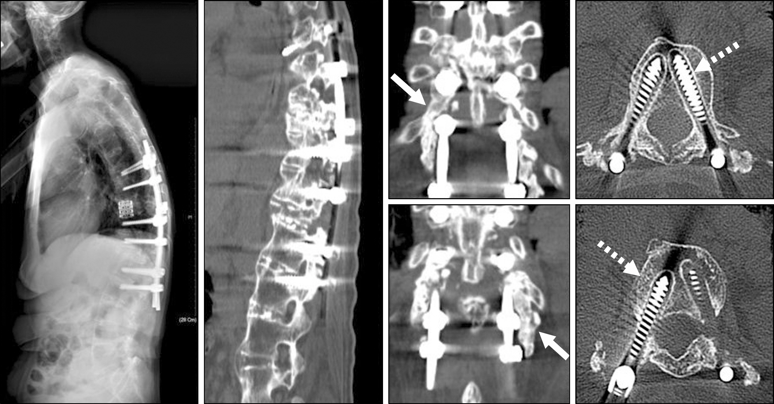
Figure 4. The computed tomography after 5 months treatment with the teriparatide demonstrated no specific interval change for the screw loosening (dot arrows). But the grafted bone was united (solid arrows) and there was no additional loss of correction.
Reference
-
References
1. Alkhiary YM, Gerstenfeld LC, Krall E, Westmore M, Sato M, Mitlak BH, et al. Enhancement of experimental fracture-healing by systemic administration of recombinant human parathyroid hormone (PTH 1–34). J Bone Joint Surg Am. 2005; 87:731–41.
Article2. Aspenberg P, Johansson T. Teriparatide improves early callus formation in distal radial fractures. Acta Orthop. 2010; 81:234–6.
Article3. Lioubavina-Hack N, Lang NP, Karring T. Significance of primary stability for osseointegration of dental implants. Clin Oral Implants Res. 2006; 17:244–50.
Article4. Baas J. Adjuvant therapies of bone graft around non-ce-mented experimental orthopedic implants stereological methods and experiments in dogs. Acta Orthop Suppl. 2008; 79:1–43.
Article5. Schatzker J, Horne JG, Sumner-Smith G. The effect of movement on the holding power of screws in bone. Clin Orthop Relat Res. 1975; (111):257–62.
Article6. Kim HJ, Kim SG, Lee HM, Kim HS, Moon ES, Park JO, et al. Risk factors associated with the halo phenomenon after lumbar fusion surgery and its clinical significance. Asian Spine J. 2008; 2:22–6.
Article7. Iolascon G, Gimigliano F, Resmini G. Teriparatide and orthopedic surgery. Aging Clin Exp Res. 2007; 19(4 Suppl):S22–5.
- Full Text Links
-
- Actions
-
Cited
- CITED
-
- Close
- Share
-
- Similar articles
-
- Efficacy of Antibiotic-Loaded Cement Augmentation for Correcting Low Grade Pedicle Screw Loosening
- “V†Shape Corrective Osteotomy of the Long Bone
- Re-stooping after Corrective Osteotomy in Patients with Ankylosing Spondylitis
- A Correction of Malunion or Deformity in the Lower Extremity
- Multi-Rod Constructs Can Increase the Incidence of Iliac Screw Loosening after Surgery for Adult Spinal Deformity
