J Korean Assoc Oral Maxillofac Surg.
2017 Aug;43(4):282-285. 10.5125/jkaoms.2017.43.4.282.
Actinomyces: a deceptive infection of oral cavity
- Affiliations
-
- 1Department of Trauma and Emergency, All India Institute of Medical Science, Bhopal, India. thukral_rishi@yahoo.co.in
- 2Department of Prosthodontics, Crown and Bridges, People's Dental Academy, Bhopal, India.
- 3Department of Oral and Maxillofacial Pathology, People's College of Dental Sciences and Research Centre, Bhopal, India.
- 4Department of Prosthodontics, Crown and Bridges, RKDF Dental College, Bhopal, India.
- KMID: 2391361
- DOI: http://doi.org/10.5125/jkaoms.2017.43.4.282
Abstract
- Actinomycosis is an infrequent chronic infection regarded as the most misdiagnosed disease by experienced clinicians. The Office of Rare Diseases at the National Institute of Health has also listed this disease as a "rare disease." This article presents a case report of actinomycosis of the alveolus with unusual clinical features but a successful resolution. It also states the importance of biopsy of deceptive inflammatory lesions that do not respond or recur after conventional treatment modalities.
Keyword
Figure
-
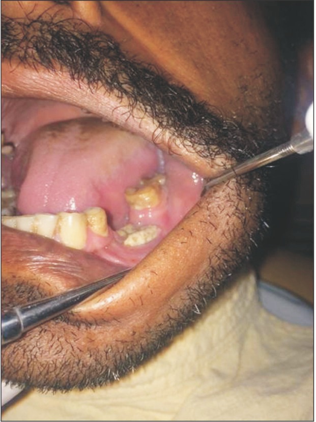
Fig. 1 Intraoral picture.
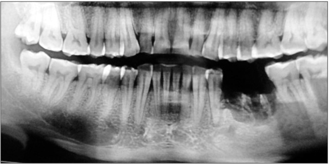
Fig. 2 Orthopantogram.
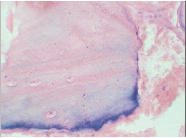
Fig. 3 Histomicrograph showing decorticated bone tissue (H&E staining, ×100).

Fig. 4 Histomicrograph revealing colonies of actinomyces (H&E staining, ×100).
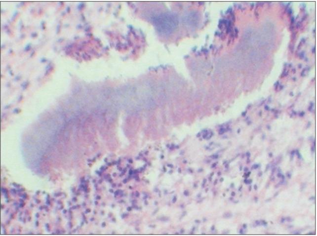
Fig. 5 Histomicrograph demonstrating the Splendore-Hoeppli phenomenon (H&E staining, ×100).
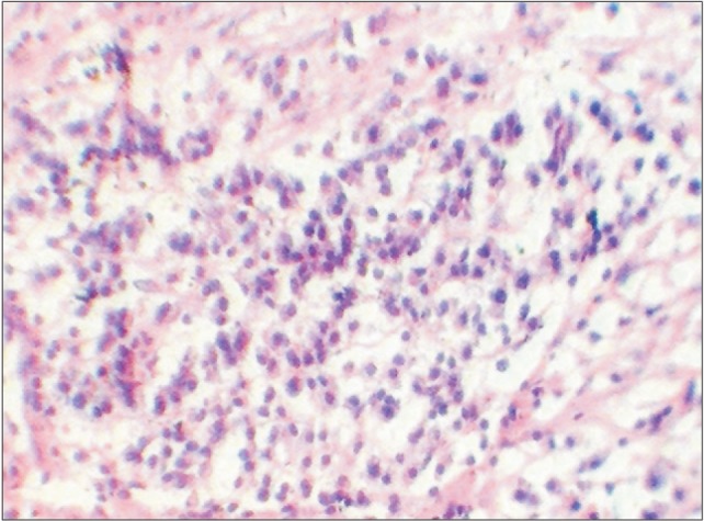
Fig. 6 Histomicrograph showing lymphoplasmocytic infiltration (H&E staining, ×100).

Fig. 7 Histomicrograph of a Gomori stain with eosin as a counter stain (×100).
Reference
-
1. Crossman T, Herold J. Actinomycosis of the maxilla: a case report of a rare oral infection presenting in general dental practice. Br Dent J. 2009; 206:201–202. PMID: 19247335.2. Boyanova L, Sabov R, Kolarov R, Mitov I. Chronic odontogenic osteomyelitis and facial actinomycosis of six month duration. JMM Case Rep. 2014; DOI: 10.1099/jmmcr.0.000729.3. Moniruddin ABM, Begum H, Nahar K. Actinomycosis: an update. Med Today. 2010; 22:43–47.
Article4. Sakallioğlu U, Açikgöz G, Kirtiloğlu T, Karagöz F. Rare lesions of the oral cavity: case report of an actinomycotic lesion limited to the gingiva. J Oral Sci. 2003; 45:39–42. PMID: 12816363.5. Rathnaprabhu V, Rajesh R, Sunitha M. Intraoral actinomycotic lesion: a case report. J Indian Soc Pedod Prev Dent. 2003; 21:144–146. PMID: 14765614.6. Valour F, Sénéchal A, Dupieux C, Karsenty J, Lustig S, Breton P, et al. Actinomycosis: etiology, clinical features, diagnosis, treatment, and management. Infect Drug Resist. 2014; 7:183–197. PMID: 25045274.7. Yadegarynia D, Merza MA, Sali S, Firuzkuhi AG. A rare case presentation of oral actinomycosis. Int J Mycobacteriol. 2013; 2:187–189. PMID: 26785990.
Article8. Gupta N, Ahmed MBR, Jadhav K. Deceptive lesion in the palate: a case report. Indian J Multidis Dent. 2013; 4:880–883.9. Behbehani MJ, Heeley JD, Jordan HV. Comparative histopathology of lesions produced by Actinomyces israelii, Actinomyces naeslundii, and Actinomyces viscosus in mice. Am J Pathol. 1983; 110:267–274. PMID: 6829706.
- Full Text Links
-
- Actions
-
Cited
- CITED
-
- Close
- Share
-
- Similar articles
-
- A Case of Esophagogastroduodenoscopy Associated Actinomycosis Presenting as Ulcers of Hard Palate
- A Rare Case of Actinomycosis in Nasal Cavity with Aspergillus Sinusitis
- Antimicrobial activity of candidate probiotic Streptococcus salivarius against Gram-positive bacteria in oral cavity
- A Case of Actinomycosis in Nasal Cavity
- A Case of Chronic Noninvasive Actinomycosis in the Nasal Cavity
