Squamous cell carcinoma arising within a maxillary odontogenic keratocyst: A rare occurrence
- Affiliations
-
- 1Eliray Oral and Maxillofacial Radiology Consulting Services, Miami, FL, USA.
- 2Department of Craniofacial Sciences, Division of Oral and Maxillofacial Surgery, University of Connecticut, School of Dental Medicine, Farmington, CT, USA.
- 3Section of Oral and Maxillofacial Radiology, University of Connecticut, School of Dental Medicine, Farmington, CT, USA. Tadinada@uchc.edu
- KMID: 2389895
- DOI: http://doi.org/10.5624/isd.2017.47.2.135
Abstract
- Squamous cell carcinoma (SCC) arising within the lining of an odontogenic keratocyst (OKC) is a rare occurrence. Although potentially locally destructive, OKC is a benign odontogenic process that typically presents with clinical and radiographic features characteristic of a benign intraosseous neoplasm. We present the clinical and radiographic features of a maxillary mass that demonstrated SCC arising from the lining of an OKC. Although the initial clinical and radiographic presentation suggested an infection or malignant neoplasm, biopsies revealed an infiltrative well-differentiated SCC contiguous with and arising from the focus of a pre-existing OKC. The patient subsequently underwent a type II hemi-maxillectomy with neoadjuvant chemoradiation. This report discusses the clinical and radiographic features associated with intraosseous malignancies, especially those arising from an otherwise benign odontogenic lesion. While the majority of OKCs are benign, the current report illustrates the potential for carcinomatous transformation within the lining of an OKC.
MeSH Terms
Figure
-
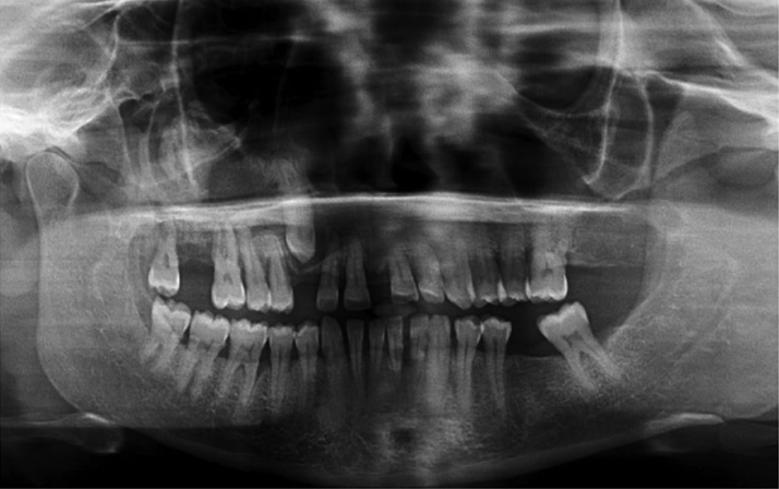
Fig. 1 Preoperative panoramic radiograph demonstrates extensive bone destruction in the right maxilla. The lesion is ill-defined, with ‘blow-out’ of much of the right antrum, also involving the right orbital floor. The maxillary canine is impacted by a pericoronal radiolucency that appears well defined.

Fig. 2 A and B. Axial bone window and coronal soft tissue algorithms of CT show the impacted right maxillary canine surrounded by a large, well defined, low intensity lesion growing more along the bone than buccal palatal expansion, extending through the palate and expanding and thinning the buccal cortical border adjacent to the right canine. C and D. Axial bone window and coronal soft tissue algorithm of CT show erosion of much of the floor, anterior and lateral walls of the right maxillary sinus, obliteration of the entire right antral cavity and invasion of the surrounding soft tissue by the lesion. The right lateral nasal wall adjacent to inferior concha and ipsilateral orbital floor are also eroded and edema of the superficial soft tissue on the right side is evident.
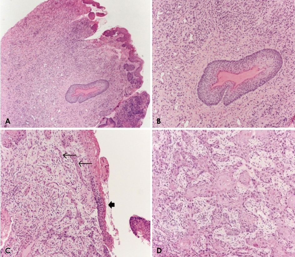
Fig. 3 Squamous cell carcinoma arising in an odontogenic keratocyst. A. Low-power magnification demonstrates the cyst lining and a satellite cyst within the wall. B. A satellite cyst demonstrates classic odontogenic keratocyst lining epithelium. Note the regimented basal cells and the uniform thickness of the epithelium. C. Odontogenic keratocyst lining demonstrates dysplastic changes. Note the nests of SCC arising from and contiguous with the cyst lining. D. A high-power view shows the infiltrative squamous cell carcinoma.
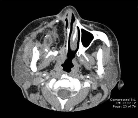
Fig. 4 A postoperative axial computed tomography scan reveals total right maxillectomy with right orbital exoneration and free flap reconstruction. No residual tumor or fluid was detected.
Reference
-
1. Maurette PE, Jorge J, de Moraes M. Conservative treatment protocol of odontogenic keratocyst: a preliminary study. J Oral Maxillofac Surg. 2006; 64:379–379.
Article2. Habibi A, Saghravanian N, Habibi M, Mellati E, Habibi M. Keratocystic odontogenic tumor: a 10-year retrospective study of 83 cases in an Iranian population. J Oral Sci. 2007; 49:229–235.
Article3. Sharif FNj, Oliver R, Sweet C, Sharif MO. Interventions for the treatment of keratocystic odontogenic tumors (KCOT, odontogenic keratocyst (OKC)). Cochrane Database Syst Rev. 2010; 9:CD008464.4. Mendes RA, Carvalho JF, Van der Waal I. Characterization and management of the keratocystic odontogenic tumor in relation to its histological and biological features. Oral Oncol. 2010; 46:219–225.5. Soskolne WA, Shear M. Observations on the pathogenesis of primordial cysts. Br Dent J. 1967; 123:321–326.6. Chirapathomsakul D, Sastravaha P, Jansisyanont P. A review of odontogenic keratocysts and the behavior of recurrences. Oral Surg Oral Med Oral Pathol Oral Radiol Endod. 2006; 101:5–10.
Article7. Shear M. The aggressive nature of the odontogenic keratocyst: is it a benign cystic neoplasm? Part 1. Clinical and early experimental evidence of aggressive behaviour. Oral Oncol. 2002; 38:219–226.
Article8. Browne RM, Gough NG. Malignant change in the epithelium of lining odontogenic cysts. Cancer. 1972; 29:1199–1207.9. Fanibunda K, Soames JV. Malignant and premalignant change in odontogenic cysts. J Oral Maxillofac Surg. 1995; 53:1469–1472.
Article10. Falaki F, Delavarian Z, Salehinejad J, Saghafi S. Squamous cell carcinoma arising from an odontogenic keratocyst: a case report. Med Oral Patol Oral Cir Bucal. 2009; 14:E171–E174.11. Lee JW, Gates R, Wignall A. Squamous cell carcinoma arising from a keratocystic odontogenic tumor. Otolaryngol Head Neck Surg. 2011; 145:356–357.
Article12. Batsakis JG, Rice DH, Solomon AR. The pathology of head and neck tumors: squamous and mucous-gland carcinomas of the nasal cavity, paranasal sinuses, and larynx, part 6. Head Neck Surg. 1980; 2:497–508.
Article13. Slootweg PJ, Richardon M. Squamous cell carcinoma of the upper aerodigestive system. In : Gnepp DR, editor. Diagnostic surgical pathology of the head and neck. 2nd ed. Philadelphia, PA: Saunders Elsevier;2009. p. 45–110.14. Syrjänen KJ. HPV infections in benign and malignant sinonasal lesions. J Clin Pathol. 2003; 56:174–181.15. Mills SE. Neuroectodermal neoplasms of the head and neck with emphasis on neuroendocrine carcinomas. Mod Pathol. 2002; 15:264–278.
Article16. Agaram NP, Collins BM, Barnes L, Lomago D, Aldeeb D, Swalsky P, et al. Molecular analysis to demonstrate that odontogenic keratocysts are neoplastic. Arch Pathol Lab Med. 2004; 128:313–317.
Article17. Gomes CC, Diniz MG, Gomez RS. Review of the molecular pathogenesis of the odontogenic keratocyst. Oral Oncol. 2009; 45:1011–1014.
Article18. Henley J, Summerlin DJ, Tomich C, Zhang S, Cheng L. Molecular evidence supporting the neoplastic nature of odontogenic keratocyst: a laser capture microdissection study of 15 cases. Histopathology. 2005; 47:582–586.
Article19. Yoshida H, Onizawa K, Yusa H. Squamous cell carcinoma arising in association with an orthokeratinized odontogenic keratocyst. Report of a case. J Oral Maxillofac Surg. 1996; 54:647–651.20. Minić AJ. Primary intraosseous squamous cell carcinoma arising in a mandibular keratocyst. Int J Oral Maxillofac Surg. 1992; 21:163–165.
Article21. Areen RG, McClatchey KD, Baker HI. Squamous cell carcinoma developing in an odontogenic keratocyst. Report of a case. Arch Otolaryngol. 1981; 107:568–569.
Article22. Makowski GJ, McGuff S, Van Sickels JE. Squamous cell carcinoma in a maxillary odontogenic keratocyst. J Oral Maxillofac Surg. 2001; 59:76–80.
Article23. Chaisuparat R, Coletti D, Kolokythas A, Ord RA, Nikitakis NG. Primary intraosseous odontogenic carcinoma arising in an odontogenic cyst or de novo: a clinicopathologic study of six new cases. Oral Surg Oral Med Oral Pathol Oral Radiol Endod. 2006; 101:194–200.
Article24. Waldron CA, Mustoe TA. Primary intraosseous carcinoma of the mandible with probable origin in an odontogenic cyst. Oral Surg Oral Med Oral Pathol. 1989; 67:716–724.
Article25. Meara JG, Shah S, Li KK, Cunningham MJ. The odontogenic keratocyst: a 20-year clinicopathologic review. Laryngoscope. 1998; 108:280–283.26. Sato T, Kamiya Y, Tochihara S, Tochihara S, Toyoda N, Asada K, et al. Primary intraosseous squamous cell carcinoma of the mandible: report of a case and review of the recent literature. Oral Med Pathol. 2006; 11:83–88.27. McDonald AR, Pogrel MA, Carson J, Regezi J. p53-positive squamous cell carcinoma originating from an odontogenic cyst. J Oral Maxillofac Surg. 1996; 54:216–218.
Article28. Anand VK, Arrowood JP Jr, Krolls SO. Malignant potential of the odontogenic keratocyst. Otolaryngol Head Neck Surg. 1994; 111:124–129.
Article29. Ward TG, Cohen B. Squamous carcinoma in a mandibular cyst. Br J Oral Surg. 1963; 1:8–12.
Article30. Bodner L, Manor E, Shear M, van der Waal I. Primary intraosseous squamous cell carcinoma arising in an odontogenic cyst: a clinicopathologic analysis of 116 reported cases. J Oral Pathol Med. 2011; 40:733–738.31. Thomas G, Pandey M, Mathew A, Abraham EK, Francis A, Somanathan T, et al. Primary intraosseous carcinoma of the jaw: pooled analysis of world literature and report of two new cases. Int J Oral Maxillofac Surg. 2001; 30:349–355.
Article32. Johnson LM, Sapp JP, Mcintre DM. Squamous cell carcinoma arising in a dentigerous cyst. J Oral Maxillofac Surg. 1994; 52:987–990.
Article
- Full Text Links
-
- Actions
-
Cited
- CITED
-
- Close
- Share
-
- Similar articles
-
- A Case of Squamous Cell Carcinoma arising from an Odontogenic Keratocyst
- Squamous cell carcinoma arising in an odontogenic cyst
- Removal of Odontogenic Keratinocyst using Versatile Maxillary Window in BCNS
- Squamous cell carcinoma arising from residual odontogenic cyst: Report of a Case & Review of Literatures
- Odontogenic Keratocyst Associated with an Ectopic Tooth in the Maxillary Sinus: A Report of Two Cases and a Review of the Literature
