Imaging Sci Dent.
2017 Jun;47(2):87-92. 10.5624/isd.2017.47.2.87.
Is the panoramic mandibular index useful for bone quality evaluation?
- Affiliations
-
- 1Department of Oral and Maxillofacial Radiology and Dental Research Institute, School of Dentistry, Seoul National University, Seoul, Korea. hmslsh@snu.ac.kr
- KMID: 2389888
- DOI: http://doi.org/10.5624/isd.2017.47.2.87
Abstract
- PURPOSE
The aim of this study was to determine whether the panoramic mandibular index (PMI) is useful for assessing bone mineral density. We also analyzed the potential correlations between PMI parameters and patient age.
MATERIALS AND METHODS
Four observers measured the PMI of both sides of the mental foramen using a picture archiving and communication system and images in the Digital Imaging and Communications in Medicine format. They studied 300 panoramic radiographic images of patients belonging to the following age groups: 40-49 years, 50-59 years, 60-69 years, 70-79 years, and 80-89 years. The observers were allowed to zoom in or out and to adjust the contrast of the images. Further, they were instructed to record the reasons for any measurements that could not be made. Then, we conducted a reliability analysis of the measured PMI and assessed the correlations between different patient age groups and the 3 parameters used for determining the PMI from the available data.
RESULTS
Among the 600 data items collected, 23 items were considered unmeasurable by at least 1 observer for the following 4 reasons: postoperative state, lesion, unidentified mental foramen, and alveolar bone loss. The intraobserver reproducibility of the measurable data was 0.611-0.752. The mandibular cortical width (MCW) decreased significantly as patient age increased.
CONCLUSION
PMI had limited usability when the margin of the mental foramen was not clear. In contrast, MCW, a parameter used for determining the PMI, had fewer drawbacks than the PMI with respect to bone mineral density measurements and exhibited a significant correlation with patient age.
Keyword
Figure
-
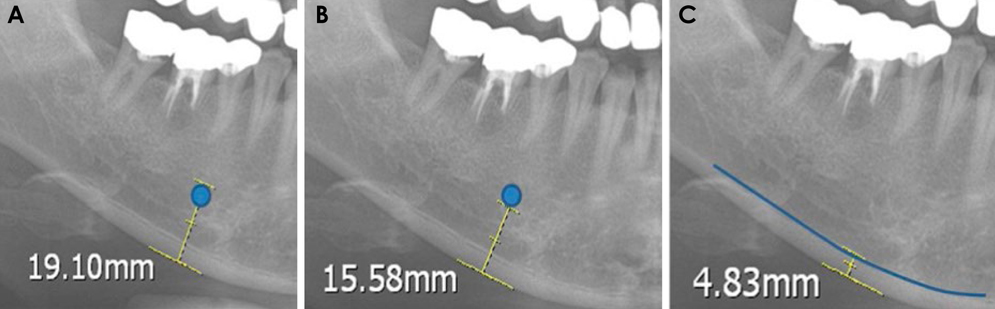
Fig. 1 A. Cropped panoramic radiograph shows the mental foramen marked with a circle and the distance measurement value. The distance between the superior margin of the mental foramen and the lower border of the mandible is 19.10 mm. B. The distance between the inferior margin of the mental foramen and the lower border of the mandible is 15.58 mm. C. The measured distance of the mandibular cortical width is 4.83 mm.
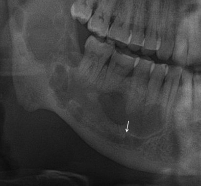
Fig. 2 The mental foramen is seen below the radiolucent lesion (arrow).
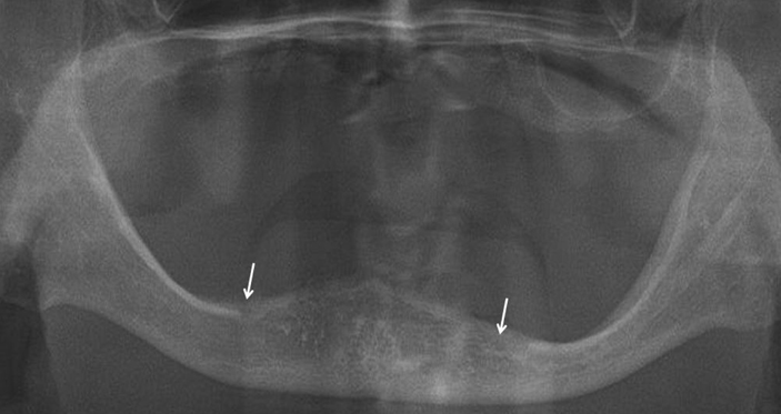
Fig. 3 A panoramic radiograph shows severe alveolar bone loss, and both sides of the mental foramen are identified near the alveolar crest level (arrow).
Reference
-
1. Assessment of fracture risk and its application to screening for postmenopausal osteoporosis. Report of a WHO Study Group. World Health Organ Tech Rep Ser. 1994; 843:1–129.2. Kanis JA. Diagnosis of osteoporosis. Osteoporos Int. 1997; 7:Suppl 3. 108–116.
Article3. Lewis MK, Blake GM, Fogelman I. Patient dose in dual X-ray absorptiometry. Osteoporos Int. 1994; 4:11–15.4. Mueller D, Gandjour A. Cost-effectiveness of using clinical risk factors with and without DXA for osteoporosis screening in postmenopausal women. Value Health. 2009; 12:1106–1117.
Article5. Devlin CV, Horner K, Devlin H. Variability in measurement of radiomorphometric indices by general dental practitioners. Dentomaxillofac Radiol. 2001; 30:120–125.
Article6. Benson BW, Prihoda TJ, Glass BJ. Variations in adult cortical bone mass as measured by a panoramic mandibular index. Oral Surg Oral Med Oral Pathol. 1991; 71:349–356.
Article7. Klemetti E, Kolmakov S, Kroger H. Pantomography in the assessment of the osteoporosis risk group. Scand J Dent Res. 1994; 102:68–72.8. Klemetti E, Kolmakov S, Heiskanen P, Vainio P, Lassila V. Panoramic mandibular index and bone mineral densities in postmenopausal women. Oral Surg Oral Med Oral Pathol. 1993; 75:774–779.
Article9. Ngeow WC, Dionysius DD, Ishak H, Nambiar P. Effect of ageing towards location and visibility of mental foramen on panoramic radiographs. Singapore Dent J. 2010; 31:15–19.
Article10. Yosue T, Brooks SL. The appearance of mental foramina on panoramic radiographs. I. Evaluation of patients. Oral Surg Oral Med Oral Pathol. 1989; 68:360–364.
Article11. Ariji Y, Katsumata A, Kubo R, Taguchi A, Fujita H, Ariji E. Factors affecting observer agreement in morphological evaluation of mandibular cortical bone on panoramic radiographs. Oral Radiol. 2017; 33:117–123.
Article12. Nakamoto T, Taguchi A, Ohtsuka M, Suei Y, Fujita M, Tanimoto K, et al. Dental panoramic radiograph as a tool to detect postmenopausal women with low bone mineral density: untrained general dental practitioners’ diagnostic performance. Osteoporos Int. 2003; 14:659–664.13. White SC, Taguchi A, Kao D, Wu S, Service SK, Yoon D, et al. Clinical and panoramic predictors of femur bone mineral density. Osteoporos Int. 2005; 16:339–346.
Article14. Alman AC, Johnson LR, Calverley DC, Grunwald GK, Lezotte DC, Hokanson JE. Diagnostic capabilities of fractal dimension and mandibular cortical width to identify men and women with decreased bone mineral density. Osteoporos Int. 2012; 23:1631–1636.
Article15. Sindeaux R, Figueiredo PT, de Melo NS, Guimaraes AT, Lazarte L, Pereira FB, et al. Fractal dimension and mandibular cortical width in normal and osteoporotic men and women. Maturitas. 2014; 77:142–148.
Article16. Roberts M, Yuan J, Graham J, Jacobs R, Devlin H. Changes in mandibular cortical width measurements with age in men and women. Osteoporos Int. 2011; 22:1915–1925.
Article17. Hastar E, Yilmaz HH, Orhan H. Evaluation of mental index, mandibular cortical index and panoramic mandibular index on dental panoramic radiographs in the elderly. Eur J Dent. 2011; 5:60–67.
Article18. Alkurt MT, Peker I, Sanal O. Assessment of repeatability and reproducibility of mental and panoramic mandibular indices on digital panoramic images. Int Dent J. 2007; 57:433–438.
Article19. Yasar F, Akgunlu F. The differences in panoramic mandibular indices and fractal dimension between patients with and without spinal osteoporosis. Dentomaxillofac Radiol. 2006; 35:1–9.20. Devlin H, Allen PD, Graham J, Jacobs R, Karayianni K, Lindh C, et al. Automated osteoporosis risk assessment by dentists: a new pathway to diagnosis. Bone. 2007; 40:835–842.
Article21. Arifin AZ, Asano A, Taguchi A, Nakamoto T, Ohtsuka M, Tsuda M, et al. Computer-aided system for measuring the mandibular cortical width on dental panoramic radiographs in identifying postmenopausal women with low bone mineral density. Osteoporos Int. 2006; 17:753–759.
Article22. Kavitha MS, Samopa F, Asano A, Taguchi A, Sanada M. Computer-aided measurement of mandibular cortical width on dental panoramic radiographs for identifying osteoporosis. J Investig Clin Dent. 2012; 3:36–44.
Article
- Full Text Links
-
- Actions
-
Cited
- CITED
-
- Close
- Share
-
- Similar articles
-
- Description of mandibular bone quality based on measurements of cortical thickness using Mental Index of male and female patients between 40-60 years old
- The correlationship between mandibular radiomorphometric indices in panorama and bone mineral density in Cu-equivalent image of intraoral film
- The relationship between age and the mandibular cortical bone thickness by using panoramic radiograph
- Comparison of panorama radiomorphometric indices of the mandible in normal and osteoporotic women
- Bone height measurements of implant sites: Comparison of panoramic radiography and spiral computed tomography
