Imaging Sci Dent.
2017 Jun;47(2):69-74. 10.5624/isd.2017.47.2.69.
Evaluation of the morphology of the canalis sinuosus using cone-beam computed tomography in patients with maxillary impacted canines
- Affiliations
-
- 1Department of Oral and Maxillofacial Surgery, Istanbul Medipol University School of Dentistry, Istanbul, Turkey. gurlergokhan@yahoo.com
- 2Department of Oral and Maxillofacial Radiology, Istanbul Medipol University School of Dentistry, Istanbul, Turkey.
- 3Department of Anatomy, Istanbul Medipol University School of Medicine, Istanbul, Turkey.
- KMID: 2389886
- DOI: http://doi.org/10.5624/isd.2017.47.2.69
Abstract
- PURPOSE
The nasopalatine canal is a well-known, important anatomical structure in the anterior maxilla, but this region contains many accessory canals. The canalis sinuosus (CS) is one of these canals; it contains the anterior superior alveolar nerve, along with veins and arteries. The purpose of this study was to evaluate the CS using conebeam computed tomography (CBCT) in patients with maxillary impacted canines.
MATERIALS AND METHODS
A total of 111 patients admitted to the Istanbul Medipol University School of Dentistry for the exposure, orthodontic treatment, and/or extraction of an impacted canine were included in this study. CBCT images were obtained for these patients under standard conditions. Axial, coronal, and sagittal sections were evaluated to assess the prevalence of CS, the direction and diameter of the canal, its relation with the impacted canine, and its distance from the alveolar crest. Further, possible correlations with patient gender and age were analyzed.
RESULTS
The CS could be detected bilaterally in all the evaluated tomography images. The mean canal diameter was significantly larger in males than in females (P=.001). The CS ran significantly closer to the impacted canine when the canal was located horizontally (P=.03). Variations of the canal, such as accessory canals, were identified in 6 patients.
CONCLUSION
CS is an anatomical entity that may resemble periapical lesions and other anatomical structures. Evaluation with CBCT prior to surgical procedures in the anterior maxilla will help to prevent overlooking such anatomical structures and to decrease possible surgical complications.
Keyword
MeSH Terms
Figure
-
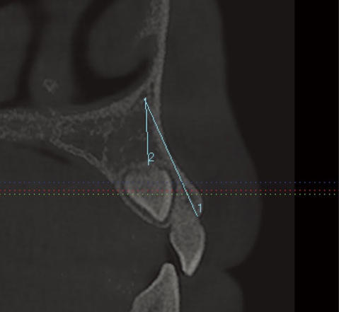
Fig. 1 The distance between the terminal portion of the canalis sinuosus (CS) and the region of the buccal alveolar ridge is measured on the sagittal plane.
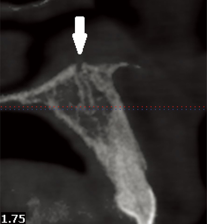
Fig. 2 The terminal portion of the CS merges superiorly with the nasopalatine canal.
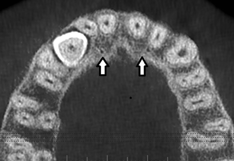
Fig. 3 Accessory bony canals run downward towards the alveolar ridge.
Reference
-
1. Wanzeler AM, Marinho CG, Alves Junior SM, Manzi FR, Tuji FM. Anatomical study of the canalis sinuosus in 100 cone beam computed tomography examinations. Oral Maxillofac Surg. 2015; 19:49–53.
Article2. von Arx T, Lozanoff S, Sendi P, Bornstein MM. Assessment of bone channels other than the nasopalatine canal in the anterior maxilla using limited cone beam computed tomography. Surg Radiol Anat. 2013; 35:783–790.
Article3. Tanaka R, Hayashi T, Ohshima H, Ida-Yonemochi H, Kenmotsu S, Ike M. CT anatomy of the anterior superior alveolar nerve canal: a macroscopic and microscopic study. Oral Radiol. 2011; 27:93–97.
Article4. Neves FS, Crusoé-Souza M, Franco LC, Caria PH, Bonfim-Almeida P, Crusoé-Rebello I. Canalis sinuosus: a rare anatomical variation. Surg Radiol Anat. 2012; 34:563–566.
Article5. Torres MG, de Faro Valverde L, Vidal MT, Crusoé-Rebello IM. Branch of the canalis sinuosus: a rare anatomical variation-a case report. Surg Radiol Anat. 2015; 37:879–881.
Article6. de Oliveira-Santos C, Rubira-Bullen IR, Monteiro SA, León JE, Jacobs R. Neurovascular anatomical variations in the anterior palate observed on CBCT images. Clin Oral Implants Res. 2013; 24:1044–1048.7. Jones FW. The anterior superior alveolar nerve and vessels. J Anat. 1939; 73:583–591.8. von Arx T, Lozanoff S. Anterior superior alveolar nerve (ASAN). Swiss Dent J. 2015; 125:1202–1209.9. Heasman PA. Clinical anatomy of the superior alveolar nerves. Br J Oral Maxillofac Surg. 1984; 22:439–447.
Article10. Robinson S, Wormald PJ. Patterns of innervation of the anterior maxilla: a cadaver study with relevance to canine fossa puncture of the maxillary sinus. Laryngoscope. 2005; 115:1785–1788.
Article11. Shelley AM, Rushton VE, Horner K. Canalis sinuosus mimicking a periapical inflammatory lesion. Br Dent J. 1999; 186:378–379.
Article12. Olenczak JB, Hui-Chou HG, Aguila DJ 3rd, Shaeffer CA, Dellon AL, Manson PN. Posttraumatic midface pain: clinical significance of the anterior superior alveolar nerve and canalis sinuosus. Ann Plast Surg. 2015; 75:543–547.13. Sekerci AE, Cantekin K, Aydinbelge M. Cone beam computed tomographic analysis of neurovascular anatomical variations other than the nasopalatine canal in the anterior maxilla in a pediatric population. Surg Radiol Anat. 2015; 37:181–186.
Article
- Full Text Links
-
- Actions
-
Cited
- CITED
-
- Close
- Share
-
- Similar articles
-
- Retrospective Analysis of Incisor Root Resorption Associated with Impacted Maxillary Canines
- Factors Influencing the Duration of Forced Eruption in Impacted Maxillary Canines
- Invasion of the canalis sinuosus by dental implants: A report of 3 cases
- Evaluation of Impacted Maxillary Canine Position Using Panoramic Radiographs and Cone-beam Computed Tomography
- Cone beam computed tomography findings of ectopic mandibular third molar in the mandibular condyle: report of a case
