Diagnostic Performance of Intravascular Ultrasound-Derived Minimal Lumen Area to Predict Functionally Significant Non-Left Main Coronary Artery Disease: a Meta-Analysis
- Affiliations
-
- 1Division of Cardiology, Busan Paik Hospital, University of Inje College of Medicine, Busan, Korea. jsjang71@gmail.com
- 2Department of Neurology, Kangdong Sacred Heart Hospital, Hallym University College of Medicine, Seoul, Korea.
- 3Division of Cardiology, Kosin University Medical Center, Busan, Korea.
- 4Department of Internal Medicine, Pusan National University Hospital, Busan, Korea.
- 5Division of Cardiology, Pusan National University Yangsan Hospital, Yangsan, Korea.
- 6Department of Cardiovascular Surgery, Busan National University Yangsan Hospital, Yangsan, Korea.
- KMID: 2389618
- DOI: http://doi.org/10.4070/kcj.2016.46.5.622
Abstract
- BACKGROUND AND OBJECTIVES
Intravascular ultrasound (IVUS)-guided percutaneous coronary intervention frequently results in unnecessary stenting due to the low positive predictive value of IVUS-derived minimal lumen area (MLA) for identification of functionally significant coronary stenosis. We appraised the diagnostic accuracy of IVUS-derived MLA compared with the fractional flow reserve (FFR) to assess intermediate coronary stenosis.
SUBJECTS AND METHODS
We searched MEDLINE and Cochrane databases for studies using IVUS and FFR methods to establish the best MLA cut-off values to predict significant non-left main coronary artery stenosis. Summary estimates were obtained using a random-effects model.
RESULTS
The 17 studies used in our analysis enrolled 3920 patients with 4267 lesions. The weighted overall mean MLA cut-off value was 2.58 mm². The pooled MLA sensitivity that predicted functionally significant coronary stenosis was 0.75 (confidence interval [CI]: 0.72 to 0.77) and the specificity was 0.66 (CI: 0.64 to 0.68). The positive likelihood ratio (LR) was 2.33 (CI: 2.06 to 2.63) and LR (-) was 0.33 (CI: 0.26 to 0.42). The pooled diagnostic odds ratio (DOR) was 7.53 (CI: 5.26 to 10.76) and the area under the summary receiver operating characteristic curve for all the trials was 0.782 with a Q point of 0.720. Meta-regression analysis demonstrated that an FFR cut-off point of 0.75 was associated with a four times higher diagnostic accuracy compared to that of 0.80 (relative DOR: 3.92; 95% CI: 1.25 to 12.34).
CONCLUSION
IVUS-derived MLA has limited diagnostic accuracy and needs careful interpretation to correlate with functionally significant non-left main coronary artery stenosis.
MeSH Terms
Figure
-

Fig. 1 Trial flow chart shows number of studies retrieved by individual searches and number of trials included in review.
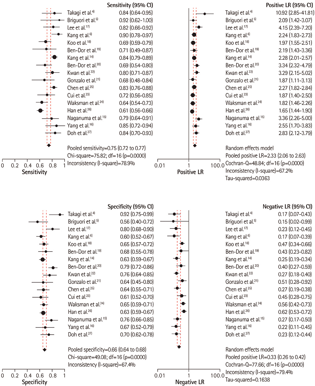
Fig. 2 Combined sensitivity, specificity, positive and negative LRs of the included trials. The MLA derived from IVUS has a limited pooled diagnostic performance in predicting functionally significant non-left main coronary artery stenosis. LR: likelihood ratio, MLA: minimal lumen area, IVUS: intravascular ultrasound, CI: confidence interval.
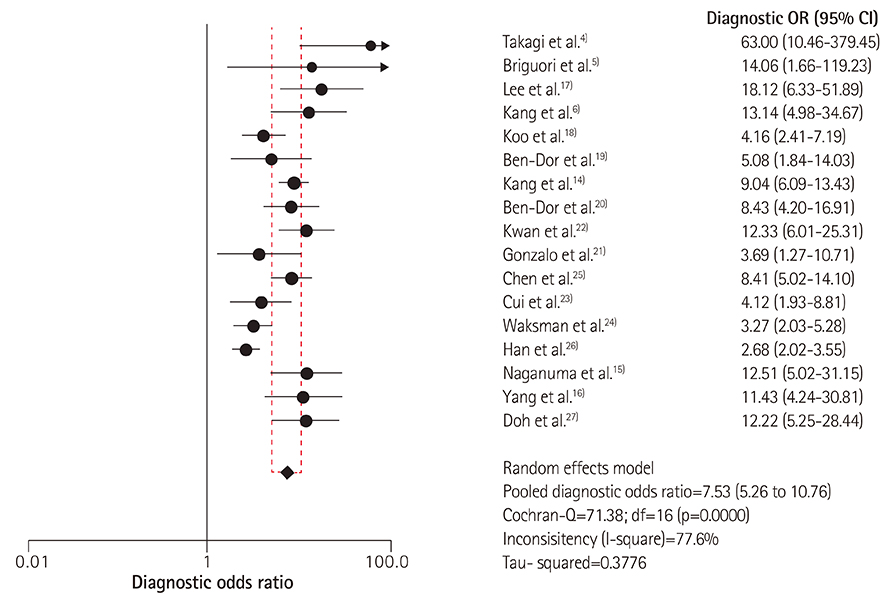
Fig. 3 Pooled DOR of the included studies. The odds of positive intravascular ultrasound results were 7.53 times higher in patients with functionally significant disease compared to the odds of positive results in patients without significant disease. DOR: diagnostic odds ratio.
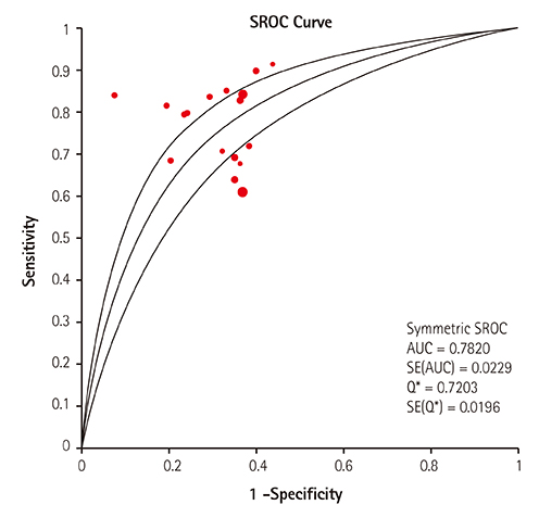
Fig. 4 Summary ROC curve of the included studies. The area under the summary ROC curve for all the trials was 0.782 with the Q point (Q*) of 0.720. The upper and lower lines indicate 95% confidence intervals. ROC: receiver operating characteristic. SROC: summary receiver operating characteristic, AUC: area under the curve, SE: standard error.
Cited by 2 articles
-
Ischemia-based Coronary Revascularization: Beyond Anatomy and Fractional Flow Reserve
Hong-Seok Lim, Kyoung-Woo Seo, Myeong-Ho Yoon, Hyoung-Mo Yang, Seung-Jea Tahk
Korean Circ J. 2018;48(1):16-23. doi: 10.4070/kcj.2017.0177.The Current Status of Intervention for Intermediate Coronary Stenosis in the Korean Percutaneous Coronary Intervention (K-PCI) Registry
Jin-Ho Kim, Woonggil Choi, Ki-Chang Kim, Chang-Wook Nam, Bum-Kee Hong, June-Hong Kim, Doo Soo Jeon, Jang-Whan Bae, Sang-Hyun Kim, Keon-Woong Moon, Byung-Ryul Cho, Doo Il Kim, Jae-Sik Jang
Korean Circ J. 2019;49(11):1022-1032. doi: 10.4070/kcj.2019.0074.
Reference
-
1. Bech GJ, De Bruyne B, Pijls NH, et al. Fractional flow reserve to determine the appropriateness of angioplasty in moderate coronary stenosis: a randomized trial. Circulation. 2001; 103:2928–2934.2. Fearon WF, Bornschein B, Tonino PA, et al. Economic evaluation of fractional flow reserve-guided percutaneous coronary intervention in patients with multivessel disease. Circulation. 2010; 122:2545–2550.3. Jang JS, Song YJ, Kang W, et al. Intravascular ultrasound-guided implantation of drug-eluting stents to improve outcome: a meta-analysis. JACC Cardiovasc Interv. 2014; 7:233–243.4. Takagi A, Tsurumi Y, Ishii Y, Suzuki K, Kawana M, Kasanuki H. Clinical potential of intravascular ultrasound for physiological assessment of coronary stenosis: relationship between quantitative ultrasound tomography and pressure-derived fractional flow reserve. Circulation. 1999; 100:250–255.5. Briguori C, Anzuini A, Airoldi F, et al. Intravascular ultrasound criteria for the assessment of the functional significance of intermediate coronary artery stenoses and comparison with fractional flow reserve. Am J Cardiol. 2001; 87:136–141.6. Kang SJ, Lee JY, Ahn JM, et al. Validation of intravascular ultrasound-derived parameters with fractional flow reserve for assessment of coronary stenosis severity. Circ Cardiovasc Interv. 2011; 4:65–71.7. Leeflang MM, Deeks JJ, Gatsonis C, Bossuyt PM. Cochrane Diagnostic Test Accuracy Working Group. Systematic reviews of diagnostic test accuracy. Ann Intern Med. 2008; 149:889–897.8. Deeks JJ, Altman DG. Diagnostic tests 4: likelihood ratios. BMJ. 2004; 329:168–169.9. Moses LE, Shapiro D, Littenberg B. Combining independent studies of a diagnostic test into a summary ROC curve: data-analytic approaches and some additional considerations. Stat Med. 1993; 12:1293–1316.10. Dinnes J, Deeks J, Kirby J, Roderick P. A methodological review of how heterogeneity has been examined in systematic reviews of diagnostic test accuracy. Health Technol Assess. 2005; 9:1–113. iii11. Jasti V, Ivan E, Yalamanchili V, Wongpraparut N, Leesar MA. Correlations between fractional flow reserve and intravascular ultrasound in patients with an ambiguous left main coronary artery stenosis. Circulation. 2004; 110:2831–2836.12. Kang SJ, Lee JY, Ahn JM, et al. Intravascular ultrasound-derived predictors for fractional flow reserve in intermediate left main disease. JACC Cardiovasc Interv. 2011; 4:1168–1174.13. Park SJ, Ahn JM, Kang SJ, et al. Intravascular ultrasound-derived minimal lumen area criteria for functionally significant left main coronary artery stenosis. JACC Cardiovasc Interv. 2014; 7:868–874.14. Kang SJ, Ahn JM, Song H, et al. Usefulness of minimal luminal coronary area determined by intravascular ultrasound to predict functional significance in stable and unstable angina pectoris. Am J Cardiol. 2012; 109:947–953.15. Naganuma T, Latib A, Costopoulos C, et al. The role of intravascular ultrasound and quantitative angiography in the functional assessment of intermediate coronary lesions: correlation with fractional flow reserve. Cardiovasc Revasc Med. 2014; 15:3–7.16. Yang HM, Tahk SJ, Lim HS, et al. Relationship between intravascular ultrasound parameters and fractional flow reserve in intermediate coronary artery stenosis of left anterior descending artery: intravascular ultrasound volumetric analysis. Catheter Cardiovasc Interv. 2014; 83:386–394.17. Lee CH, Tai BC, Soon CY, et al. New set of intravascular ultrasound-derived anatomic criteria for defining functionally significant stenoses in small coronary arteries (results from Intravascular Ultrasound Diagnostic Evaluation of Atherosclerosis in Singapore [IDEAS] study). Am J Cardiol. 2010; 105:1378–1384.18. Koo BK, Yang HM, Doh JH, et al. Optimal intravascular ultrasound criteria and their accuracy for defining the functional significance of intermediate coronary stenoses of different locations. JACC Cardiovasc Interv. 2011; 4:803–811.19. Ben-Dor I, Torguson R, Gaglia MA Jr, et al. Correlation between fractional flow reserve and intravascular ultrasound lumen area in intermediate coronary artery stenosis. EuroIntervention. 2011; 7:225–233.20. Ben-Dor I, Torguson R, Deksissa T, et al. Intravascular ultrasound lumen area parameters for assessment of physiological ischemia by fractional flow reserve in intermediate coronary artery stenosis. Cardiovasc Revasc Med. 2012; 13:177–182.21. Gonzalo N, Escaned J, Alfonso F, et al. Morphometric assessment of coronary stenosis relevance with optical coherence tomography: a comparison with fractional flow reserve and intravascular ultrasound. J Am Coll Cardiol. 2012; 59:1080–1089.22. Kwan TW, Yang S, Xu B, et al. Optimized quantitative angiographic and intravascular ultrasound parameters predicting the functional significance of single de novo lesions in the left anterior descending artery. Chin Med J (Engl). 2012; 125:4249–4253.23. Cui M, Zhu D, Guo LJ, et al. Usefulness of lumen area parameters determined by intravascular ultrasound to predict functional significance of intermediate coronary artery stenosis. Chin Med J (Engl). 2013; 126:1606–1611.24. Waksman R, Legutko J, Singh J, et al. FIRST: Fractional flow reserve and intravascular ultrasound relationship study. J Am Coll Cardiol. 2013; 61:917–923.25. Chen SL, Xu B, Chen JB, et al. Diagnostic accuracy of quantitative angiographic and intravascular ultrasound parameters predicting the functional significance of single de novo lesions. Int J Cardiol. 2013; 168:1364–1369.26. Han JK, Koo BK, Park KW, et al. Optimal intravascular ultrasound criteria for defining the functional significance of intermediate coronary stenosis: an international multicenter study. Cardiology. 2014; 127:256–262.27. Doh JH, Koo BK, Nam CW, et al. Diagnostic value of coronary CT angiography in comparison with invasive coronary angiography and intravascular ultrasound in patients with intermediate coronary artery stenosis: results from the prospective multicentre FIGURE-OUT (Functional Imaging criteria for GUiding REview of invasive coronary angiOgraphy, intravascular Ultrasound, and coronary computed Tomographic angiography) study. Eur Heart J Cardiovasc Imaging. 2014; 15:870–877.28. Pijls NH, De Bruyne B, Peels K, et al. Measurement of fractional flow reserve to assess the functional severity of coronary-artery stenoses. N Engl J Med. 1996; 334:1703–1708.29. Nascimento BR, de Sousa MR, Koo BK, et al. Diagnostic accuracy of intravascular ultrasound-derived minimal lumen area compared with fractional flow reserve--meta-analysis: pooled accuracy of IVUS luminal area versus FFR. Catheter Cardiovasc Interv. 2014; 84:377–385.30. Tonino PA, De Bruyne B, Pijls NH, et al. Fractional flow reserve versus angiography for guiding percutaneous coronary intervention. N Engl J Med. 2009; 360:213–224.
- Full Text Links
-
- Actions
-
Cited
- CITED
-
- Close
- Share
-
- Similar articles
-
- Erratum: Diagnostic Performance of Intravascular Ultrasound-Derived Minimal Lumen Area to Predict Functionally Significant Non-Left Main Coronary Artery Disease: a Meta-Analysis
- The Relationship Between Body Mass Index and Left Main Coronary Artery Architecture: A Virtual Histology Intravascular Ultrasound Analysis
- Deep Learning-Based Lumen and Vessel Segmentation of Intravascular Ultrasound Images in Coronary Artery Disease
- Past, Present, and Future of Left Main Coronary Artery PCI
- Efficacy of Interavascular Ultrasound in the Palmaz-Schatz Stent Implantation: Clinical Experience of 3 Coronary Artery Disease Patients
