Korean Circ J.
2024 Jan;54(1):30-39. 10.4070/kcj.2023.0166.
Deep Learning-Based Lumen and Vessel Segmentation of Intravascular Ultrasound Images in Coronary Artery Disease
- Affiliations
-
- 1Biomedical Engineering Research Center, Asan Institute for Life Sciences, Seoul, Korea
- 2Department of Cardiology, Asan Medical Center, University of Ulsan College of Medicine, Seoul, Korea
- KMID: 2550605
- DOI: http://doi.org/10.4070/kcj.2023.0166
Abstract
- Background and Objectives
Intravascular ultrasound (IVUS) evaluation of coronary artery morphology is based on the lumen and vessel segmentation. This study aimed to develop an automatic segmentation algorithm and validate the performances for measuring quantitative IVUS parameters.
Methods
A total of 1,063 patients were randomly assigned, with a ratio of 4:1 to the training and test sets. The independent data set of 111 IVUS pullbacks was obtained to assess the vessel-level performance. The lumen and external elastic membrane (EEM) boundaries were labeled manually in every IVUS frame with a 0.2-mm interval. The Efficient-UNet was utilized for the automatic segmentation of IVUS images.
Results
At the frame-level, Efficient-UNet showed a high dice similarity coefficient (DSC, 0.93±0.05) and Jaccard index (JI, 0.87±0.08) for lumen segmentation, and demonstrated a high DSC (0.97±0.03) and JI (0.94±0.04) for EEM segmentation. At the vessel-level, there were close correlations between model-derived vs. experts-measured IVUS parameters; minimal lumen image area (r=0.92), EEM area (r=0.88), lumen volume (r=0.99) and plaque volume (r=0.95). The agreement between model-derived vs. expert-measured minimal lumen area was similarly excellent compared to the experts' agreement. The model-based lumen and EEM segmentation for a 20-mm lesion segment required 13.2 seconds, whereas manual segmentation with a 0.2-mm interval by an expert took 187.5 minutes on average.
Conclusions
The deep learning models can accurately and quickly delineate vascular geometry. The artificial intelligence-based methodology may support clinicians’ decisionmaking by real-time application in the catheterization laboratory.
Figure
-

Figure 1 Workflow for developing the Efficient-UNet for IVUS segmentation.IVUS = intravascular ultrasound.
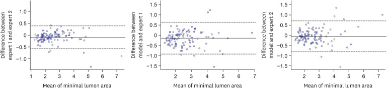
Figure 2 Bland-Altman between the proposed model-derived and expert-measured minimal lumen area.
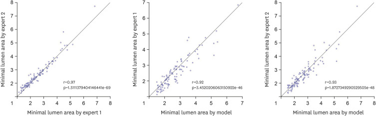
Figure 3 Correlation between the proposed model-derived and expert-measured minimal lumen area.
Reference
-
1. Fujii K, Carlier SG, Mintz GS, et al. Stent underexpansion and residual reference segment stenosis are related to stent thrombosis after sirolimus-eluting stent implantation: an intravascular ultrasound study. J Am Coll Cardiol. 2005; 45:995–998. PMID: 15808753.2. Okabe T, Mintz GS, Buch AN, et al. Intravascular ultrasound parameters associated with stent thrombosis after drug-eluting stent deployment. Am J Cardiol. 2007; 100:615–620. PMID: 17697816.3. Liu X, Doi H, Maehara A, et al. A volumetric intravascular ultrasound comparison of early drug-eluting stent thrombosis versus restenosis. JACC Cardiovasc Interv. 2009; 2:428–434. PMID: 19463466.4. Stone GW, Maehara A, Lansky AJ, et al. A prospective natural-history study of coronary atherosclerosis. N Engl J Med. 2011; 364:226–235. PMID: 21247313.5. Bajaj R, Huang X, Kilic Y, et al. Advanced deep learning methodology for accurate, real-time segmentation of high-resolution intravascular ultrasound images. Int J Cardiol. 2021; 339:185–191. PMID: 34153412.6. Zhu F, Gao Z, Zhao C, et al. A deep learning-based method to extract lumen and media-adventitia in intravascular ultrasound images. Ultrason Imaging. 2022; 44:191–203. PMID: 35861418.7. Szarski M, Chauhan S. Improved real-time segmentation of intravascular ultrasound images using coordinate-aware fully convolutional networks. Comput Med Imaging Graph. 2021; 91:101955. PMID: 34252744.8. Destrempes F, Roy Cardinal MH, Allard L, Tardif JC, Cloutier G. Segmentation method of intravascular ultrasound images of human coronary arteries. Comput Med Imaging Graph. 2014; 38:91–103. PMID: 24119335.9. Brunenberg E, Pujol O, ter Haar Romeny B, Radeva P. Automatic IVUS segmentation of atherosclerotic plaque with stop & go snake. Med Image Comput Comput Assist Interv. 2006; 9:9–16.10. Mendizabal-Ruiz EG, Rivera M, Kakadiaris IA. Segmentation of the luminal border in intravascular ultrasound B-mode images using a probabilistic approach. Med Image Anal. 2013; 17:649–670. PMID: 23490618.11. Yang J, Faraji M, Basu A. Robust segmentation of arterial walls in intravascular ultrasound images using Dual Path U-Net. Ultrasonics. 2019; 96:24–33. PMID: 30947071.12. Nishi T, Yamashita R, Imura S, et al. Deep learning-based intravascular ultrasound segmentation for the assessment of coronary artery disease. Int J Cardiol. 2021; 333:55–59. PMID: 33741429.
- Full Text Links
-
- Actions
-
Cited
- CITED
-
- Close
- Share
-
- Similar articles
-
- Deep Learning-Based Intravascular Ultrasound Images Segmentation in Coronary Artery Disease: A Start Developing the Cornerstone
- Efficacy of Interavascular Ultrasound in the Palmaz-Schatz Stent Implantation: Clinical Experience of 3 Coronary Artery Disease Patients
- Deep Learning in Dental Radiographic Imaging
- A Case of Coronary Pseudostenosis, Diagnosed by Intravascular Ultrasound
- Detection and Weak Segmentation of Masses in Gray-Scale Breast Mammogram Images Using Deep Learning
