A New Instrument for Measuring Tibial Torsion in Pediatric Patients
- Affiliations
-
- 1Department of Rehabilitation Medicine and Christian Medical Research Center, Presbyterian Medical Center, Seonam University College of Medicine, Jeonju, Korea. gvcdr@hanmail.net
- KMID: 2389460
- DOI: http://doi.org/10.5535/arm.2017.41.3.441
Abstract
OBJECTIVE
To develop and test the validity and reliability of a new instrument for measuring the thigh-foot angle (TFA) for the patients with in-toeing and out-toeing gait.
METHODS
The new instrument (Thigh-Foot Supporter [TFS]) was developed by measuring the TFA during regular examination of the tibial torsional status. The study included 40 children who presented with in-toeing and out-toeing gaits. We took a picture of each case to measure photographic-TFA (P-TFA) in the proper position and to establish a criterion. Study participants were examined by three independent physicians (A, B, and C) who had one, three and ten years of experience in the field, respectively. Each examiner conducted a separate classical physical examination (CPE) of every participant using a gait goniometer followed by a TFA assessment of each pediatric patient with or without the TFS. Thirty minutes later, repeated in the same way was measured.
RESULTS
Less experienced examiner A showed significant differences between the TFA values depending on whether TFS used (left p=0.003 and right p=0.008). However, experienced examiners B and C did not show significant differences. Using TFS, less experienced examiner A showed a high validity and all examiner's inter-test and the inter-personal reliabilities increased.
CONCLUSION
TFS may increase validity and reliability in measuring tibial torsion in patients who has a rotational problem in lower extremities. It would be more useful in less experienced examiners.
MeSH Terms
Figure
-
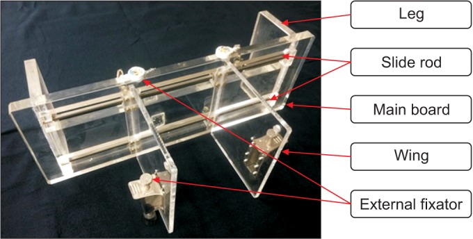
Fig. 1 Thigh-Foot Supporter.
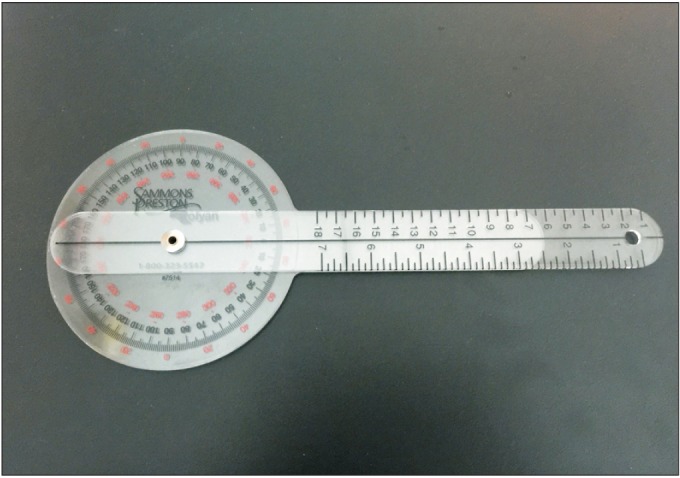
Fig. 2 Standard universal goniometer.
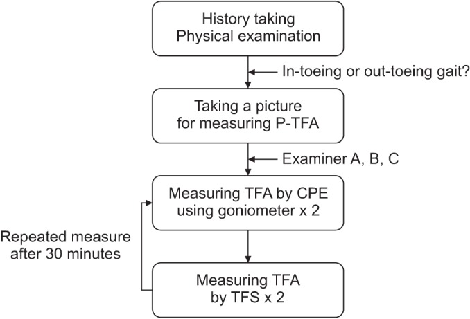
Fig. 3 Flow chart of clinical measurement. P-TFA, photographic-thigh-foot angle; CPE, classical physical examination; TFS, Thigh-Foot Supporter.
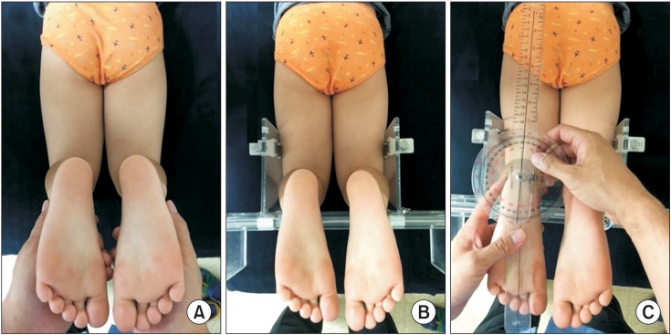
Fig. 4 Measurement of photographic thigh-foot angle (P-TFA) and thigh-foot angle (TFA) using Thigh-Foot Supporter (TFS). Lying in the prone position keeping knee joint at 90°, the ankle joint in neutral, the sole of the foot in the parallel to the floor. Then we took photos to show both thighs and feet (A). TFA was measured fixing the tibia at 90° in the sagittal and transverse plane with moving the wing of TFS (B, C).
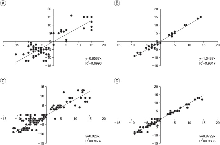
Fig. 5 Inter-test reliability of examiner A and inter-personal reliability. Inter-test reliability: (A) TFA by CPE of examiner A and (B) TFA by TFS of examiner A. Inter-personal reliability: (C) TFA by CPE and (D) TFA by TFS. TFA, thigh-foot angle; CPE, classical physical examination; TFS, Thigh-Foot Supporter.
Reference
-
1. Jang SH, Woo BS, Park SB, Lee SG. Relationship between femoral anteversion and tibial torsion in intoeing gait. J Korean Acad Rehabil Med. 1999; 23:390–396.2. Kumar SJ, MacEwen GD. Torsional abnormalities in children's lower extremities. Orthop Clin North Am. 1982; 13:629–639. PMID: 7099592.
Article3. Sass P, Hassan G. Lower extremity abnormalities in children. Am Fam Physician. 2003; 68:461–468. PMID: 12924829.4. Fabry G, Cheng LX, Molenaers G. Normal and abnormal torsional development in children. Clin Orthop Relat Res. 1994; (302):22–26.
Article5. Butler-Manuel PA, Guy RL, Heatley FW. Measurement of tibial torsion: a new technique applicable to ultrasound and computed tomography. Br J Radiol. 1992; 65:119–126. PMID: 1540801.6. Clementz BG, Magnusson A. Assessment of tibial torsion employing fluoroscopy, computed tomography and the cryosectioning technique. Acta Radiol. 1989; 30:75–80. PMID: 2914121.
Article7. Jakob RP, Haertel M, Stussi E. Tibial torsion calculated by computerized tomography and compared to other methods of measurement. J Bone Joint Surg Br. 1980; 62-B:238–242. PMID: 7364840.8. Staheli LT, Corbett M, Wyss C, King H. Lower-extremity rotational problems in children. Normal values to guide management. J Bone Joint Surg Am. 1985; 67:39–47. PMID: 3968103.
Article9. Rethlefsen SA, Healy BS, Wren TA, Skaggs DL, Kay RM. Causes of intoeing gait in children with cerebral palsy. J Bone Joint Surg Am. 2006; 88:2175–2180. PMID: 17015594.
Article10. Li YH, Leong JC. Intoeing gait in children. Hong Kong Med J. 1999; 5:360–366. PMID: 10870163.11. Kim SI, Park SB, Lee KM. Measurement of femoral anteversion using 3 dimensional imaging technique. J Korean Soc Picture Archiving Commun Syst. 1995; 1:53–58.12. Arnold AS, Komattu AV, Delp SL. Internal rotation gait: a compensatory mechanism to restore abduction capacity decreased by bone deformity. Dev Med Child Neurol. 1997; 39:40–44. PMID: 9003728.
Article13. Staheli LT. Rotational problems in children. Instr Course Lect. 1994; 43:199–209. PMID: 9097150.
Article14. Lincoln TL, Suen PW. Common rotational variations in children. J Am Acad Orthop Surg. 2003; 11:312–320. PMID: 14565753.
Article15. Kim SS, Kim SK. Derotational osteotomy and external fixation for increased femoral anteversion. J Korean Orthop Assoc. 2004; 39:361–365.
Article16. Kwon OY, Tuttle LJ, Commean PK, Mueller MJ. Reliability and validity of measures of hammer toe deformity angle and tibial torsion. Foot (Edinb). 2009; 19:149–155. PMID: 20161156.
Article17. Hernandez RJ, Tachdjian MO, Poznanski AK, Dias LS. CT determination of femoral torsion. AJR Am J Roentgenol. 1981; 137:97–101. PMID: 6787898.
Article18. Kitaoka HB, Weiner DS, Cook AJ, Hoyt WA Jr, Askew MJ. Relationship between femoral anteversion and osteoarthritis of the hip. J Pediatr Orthop. 1989; 9:396–404. PMID: 2732318.
Article19. Murphy SB, Simon SR, Kijewski PK, Wilkinson RH, Griscom NT. Femoral anteversion. J Bone Joint Surg Am. 1987; 69:1169–1176. PMID: 3667647.
Article20. Staheli LT. In-toeing and out-toeing in children. J Fam Pract. 1983; 16:1005–1011. PMID: 6842143.21. Lee SH, Chung CY, Park MS, Choi IH, Cho TJ. Tibial torsion in cerebral palsy: validity and reliability of measurement. Clin Orthop Relat Res. 2009; 467:2098–2104. PMID: 19159112.
Article22. Milner CE, Soames RW. A comparison of four in vivo methods of measuring tibial torsion. J Anat. 1998; 193(Pt 1):139–144. PMID: 9758144.
Article23. Kim HD, Lee DS, Eom MJ, Hwang JS, Han NM, Jo GY. Relationship between physical examinations and two-dimensional computed tomographic findings in children with intoeing gait. Ann Rehabil Med. 2011; 35:491–498. PMID: 22506164.
Article24. Fabry G. Normal and abnormal torsional development of the lower extremities. Acta Orthop Belg. 1997; 63:229–232. PMID: 9479773.25. Chung CY, Choi IH, Lee DY, Yoon KS, Lee DH, Sohn CS. Steel's gluteus medius and minimus advancement for in-toeing in spastic cerebral palsy. J Korean Orthop Assoc. 1996; 31:27–32.
Article
- Full Text Links
-
- Actions
-
Cited
- CITED
-
- Close
- Share
-
- Similar articles
-
- Usefulness of tibia counter rotator (TCR) for treatment of tibial internal torsion in children
- Relationship between Femoral Anteversion and Tibial Torsion in Intoeing Gait
- Effect of the Tibia Counter Rotator Orthosis for Tibial Internal Torsion Children: A Preliminary Study
- The Availability of Radiological Measurement of Tibial Torsion: Three-Dimensional Computed Tomography Reconstruction
- Tibial Torsion in Children of the Jeju Area
