The Availability of Radiological Measurement of Tibial Torsion: Three-Dimensional Computed Tomography Reconstruction
- Affiliations
-
- 1Department of Rehabilitation Medicine and Institute of Health Sciences, Gyeongsang National University Graduate School of Medicine, Jinju 660-702, Korea. hsshin@nongae.gsnu.ac.kr
- KMID: 2267229
- DOI: http://doi.org/10.5535/arm.2011.35.5.673
Abstract
OBJECTIVE
To assess the intra-rater and inter-rater reliability for measuring tibial torsion measurements by a radiographic method using three-dimensional computed tomography reconstruction (3D-CT) and to compare the physical measures to those of 3D-CT. METHOD: The study included 33 children who presented with intoeing gait. Tibial torsion was measured by 3D-CT. Distal reference point was the bimalleolar axis. Proximal reference points were the transtibial axis and posterior condylar axis. Physical measurements included thigh-foot angle (TFA) and bimalleolar angle (BMA). 3D-CT measurement and physical measurement were performed twice at both lower extremities by each rater. The intra-rater and inter-rater reliability were calculated by intraclass correlation coefficiency (ICC). The relationship between radiological and physical examination was calculated by Spearman correlation coefficient.
RESULTS
The 3D-CT measures for tibial torsion were reliable within individual raters and between different raters. However, physical measures for tibial torsion were reliable within an individual rater but not reliable between raters. The 3D-CT measures by any proximal reference axis were more reliable within a rater and between raters than physical measurements. There was no significant impact introduced by the selection of the proximal reference axis. The correlation coefficiency between 3D-CT and physical measurement methods was low.
CONCLUSION
Because the 3D-CT measurements for tibial torsion are more reliable than physical measurements, we recommend that accurate diagnosis of internal tibial torsion should be detected by using 3D-CT measurements. Also, considering the disadvantages of radiological measurements, physical measurement may be used for short term follow-up by same raters, as intra-rater reliability is relatively good.
MeSH Terms
Figure
-
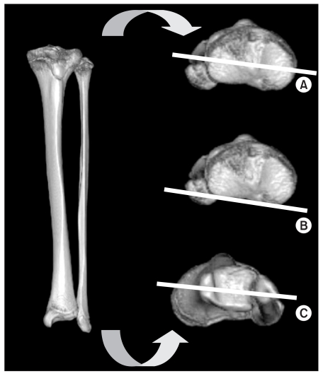
Fig. 1 Reference axes of 3-Dimensional CT reconstruction measurement. (A) Proximal transtibial axis, (B) Proximal posterior condylar axis, (C) Distal bimalleolar axis. Tibial rotation is the angle between the proximal and distal axis.
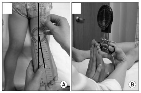
Fig. 2 Thigh foot angle which measures angle between longitudinal axis of the thigh and longitudinal axis of the foot (A) and bimalleolar angle measured by vertical goniometer at the center between medial and lateral malleolus (B), are shown.
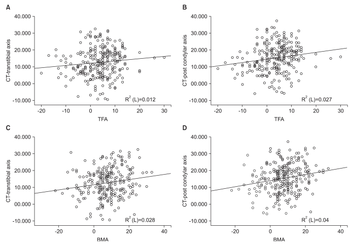
Fig. 3 Relationship between 3D-CT measurment referenced by transtibial axis as proximal axis and physical examination (TFA and BMA) (A and C) Relationship between 3D-CT measurment referenced by posterior condylar axis as proximal axis and physical examination (TFA and BMA) (B and D).
Reference
-
1. Kumar SJ, MacEwen GD. Torsional abnormalities in children's lower extremities. Orthop Clin North Am. 1982; 13:629–639. PMID: 7099592.
Article2. Staheli LT, Corbett M, Wyss C, King H. Lower-ex-tremity rotational problems in children. Normal values to guide management. J Bone Joint Surg Am. 1985; 67:39–47. PMID: 3968103.
Article3. Eckhoff DG, Johnson KK. Three-dimensional computed tomography reconstruction of tibial torsion. Clin Orthop Relat Res. 1994; 302:42–46. PMID: 8168320.
Article4. Lee S, Choi KS, Jeung IS, Lee JE, Yang SM, Lee SM. Physical examination and computed tomography in children with toe-in gait. J Korean Acad Rehabil Med. 2011; 35:61–66.5. Stuberg W, Temme J, Kaplan P, Clarke A, Fuchs R. Measurement of tibial torsion and thigh-foot angle using goniometry and computed tomography. Clin Orthop Relat Res. 1991; 272:208–212. PMID: 1934735.
Article6. Valmassy R, Stanton B. Tibial torsion. Normal values in children. J Am Podiatr Med Assoc. 1989; 79:432–435. PMID: 2778678.
Article7. Milner CE, Soames RW. A comparison of four in vivo methods of measuring tibial torsion. J Anat. 1998; 193:139–144. PMID: 9758144.
Article8. Jakob RP, Haertel M, Stussi E. Tibial torsion calculated by computerised tomography and compared to other methods of measurement. J Bone Joint Surg Br. 1980; 62:238–242. PMID: 7364840.
Article9. Lee SH, Chung CY, Park MS, Choi IH, Cho TJ. Tibial torsion in cerebral palsy: validity and reliability of measurement. Clin Orthop Relat Res. 2009; 467:2098–2104. PMID: 19159112.
Article10. Winter WG Jr, Lafferty JF. The skiing sequelae of tibial torsion. Orthop Clin North Am. 1976; 7:231–240. PMID: 1256791.
Article11. Clementz BG. Assessment of tibial torsion and rotational deformity with a new fluoroscopic technique. Clin Orthop Relat Res. 1989; 245:199–209. PMID: 2752622.
Article12. Guven M, Akman B, Unay K, Ozturan EK, Cakici H, Eren A. A new radiographic measurement method for evaluation of tibial torsion: a pilot study in adults. Clin Orthop Relat Res. 2009; 467:1807–1812. PMID: 19052824.13. Jend HH, Heller M, Dallek M, Schoettle H. Measurement of tibial torsion by computer tomography. Acta Radiol Diagn (Stockh). 1981; 22:271–276. PMID: 7315504.
Article14. Abel MF, Sutherland DH, Wenger DR, Mubarak SJ. Evaluation of CT scans and 3-D reformatted images for quantitative assessment of the hip. J Pediatr Orthop. 1994; 14:48–53. PMID: 8113372.
Article15. Kim SI, Park SB, Lee KM. Measurement of femoral anteversion using 3 dimensional imaging. J Korean Acad Rehabil Med. 1994; 18:495–499.
- Full Text Links
-
- Actions
-
Cited
- CITED
-
- Close
- Share
-
- Similar articles
-
- Relationship between Femoral Anteversion and Tibial Torsion in Intoeing Gait
- Effect of the Tibia Counter Rotator Orthosis for Tibial Internal Torsion Children: A Preliminary Study
- CT Findings of Primary Torsion of the Greater Omentum with Segmental Infarction: Case Report
- Physical Examination and Computed Tomography in Children with Toe in Gait
- The Availability of Radiological Measurement of Femoral Anteversion Angle: Three-Dimensional Computed Tomography Reconstruction
