Ultrasonography Detected Missed Lunate Volar Dislocation Associated With Median Neuropathy: A Case Report
- Affiliations
-
- 1Department of Rehabilitation Medicine, Incheon St. Mary's Hospital, College of Medicine, The Catholic University of Korea, Incheon, Korea. minukkim@nate.com
- KMID: 2389419
- DOI: http://doi.org/10.5535/arm.2017.41.4.709
Abstract
- Lunate and perilunate dislocations are uncommon, but they have clinical importance because complications, such as median neuropathy and avascular necrosis of the lunate, can occur. Although early diagnosis enabling early surgical treatment is crucial for preventing long-term sequelae, these dislocations are frequently missed in the initial assessment. Imaging tools, such as plain radiography, magnetic resonance imaging, ultrasonography, and electrodiagnostic studies, have been used for diagnosis. The proper choice of initial evaluation tools is important for making an accurate early diagnosis. Here we present a case of lunate dislocation associated with median neuropathy in which ultrasonography, along with the electrodiagnostic study and plain radiography, played an important diagnostic role in detecting structural abnormalities. This case report reveals the complementary diagnostic role of ultrasonography in initial assessment and provides ultrasonographic images of lunate dislocation as a cause of median neuropathy.
Keyword
MeSH Terms
Figure
-

Fig. 1 Wrist ultrasound images of the patient (A, B) and a normal subject (C, D). (A) A transverse 12–5 MHz ultrasound image of the patient demonstrates the median nerve and only one flexor tendon over the dislocated lunate bone. (B) A longitudinal ultrasound image of the patient clearly shows volar dislocation of the lunate bone. (C, D) Ultrasound images of a normal subject show a normal alignment of the carpal bones and a non-compressed median nerve. (C) Transverse view. (D) Longitudinal view. Sca, scaphoid; Lun, lunate; Pis, pisiform; Cap, capitate; Rad, radius; white arrow, median nerve.
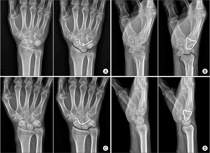
Fig. 2 Plain wrist radiographs of the patient on the symptomatic side (A, B) and the asymptomatic side (C, D). (A) The anterior-posterior view shows a triangular or wedge-shaped lunate bone and a styloid process fracture. In the right image, the ‘arcs of Gilula’, representing the carpal row borders, are illustrated. The black line marks the proximal margin of the proximal carpal row (scaphoid, lunate, and triquetrum). The gray line marks the distal margin of the same three bones. The white line marks the proximal margin of the hamate and capitate bones. The arcs of Gilula are obviously disrupted. (B) The lateral radiograph shows loss of lunate (gray line) and radius bone (black line) collinearity, with the lunate bone tilted volarly and dislocated (the ‘spilled teacup’ sign). The capitate bone is outlined in white. (C, D) Plain anterior-posterior and lateral radiographs of the asymptomatic side show normal arcs of Gilula and alignment of radius, lunate, and capitate bones. The images in (C) and (D) are flipped over the right to the left.
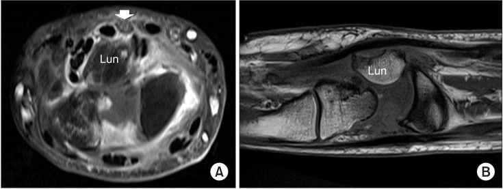
Fig. 3 Contrast-enhanced magnetic resonance imaging (A, transverse; B, coronal) additionally shows flexor tendon synovitis.

Fig. 4 Ultrasound images (A, transverse; B, longitudinal) and plain radiographs (C, anteriorposterior; D, lateral) at the wrist 1-year post-operation show good carpal bone alignment and a noncompressed median nerve (white arrow).
Reference
-
1. Youssef B, Deshmukh SC. Volar perilunate dislocation: a case report and review of the literature. Open Orthop J. 2008; 2:57–58. PMID: 19478928.
Article2. Ott F, Mattiassich G, Kaulfersch C, Ortmaier R. Initially unrecognised lunate dislocation as a cause of carpal tunnel syndrome. BMJ Case Rep. 2013; 2013.
Article3. Mayfield JK, Johnson RP, Kilcoyne RK. Carpal dislocations: pathomechanics and progressive perilunar instability. J Hand Surg Am. 1980; 5:226–241. PMID: 7400560.
Article4. Cara J, Narvaez A, de la Varga V, Guerado E. Median nerve neuropathy from an old lunate dislocation. Acta Orthop Belg. 1998; 64:100–103. PMID: 9586259.5. Sochart DH, Birdsall PD, Paul AS. Perilunate fracturedislocation: a continually missed injury. J Accid Emerg Med. 1996; 13:213–216. PMID: 8733667.
Article6. Chen WS. Median-nerve neuropathy associated with chronic anterior dislocation of the lunate. J Bone Joint Surg Am. 1995; 77:1853–1857. PMID: 8550653.
Article7. Stanbury SJ, Elfar JC. Perilunate dislocation and perilunate fracture-dislocation. J Am Acad Orthop Surg. 2011; 19:554–562. PMID: 21885701.
Article8. Peh WC, Gilula LA. Normal disruption of carpal arcs. J Hand Surg Am. 1996; 21:561–566. PMID: 8842944.
Article
- Full Text Links
-
- Actions
-
Cited
- CITED
-
- Close
- Share
-
- Similar articles
-
- Multiple Flexor Tendon Ruptures with Compression Neuropathy at Neglected Volar Lunate Dislocation
- The Treatment of Volar Iunate Dislocation and Perilunar Dislocation
- Treatment of Anchor Suture with Kirschner Wires Fixation for Chronic Perilunate Dislocation
- Carpal Tunnel Syndrome and Rupture of Flexor Tendon Associated from Neglected Anterior Lunate Dislocation
- Volar Perilunate Dislocation: A case report
