Stability of dental, alveolar, and skeletal changes after miniscrew-assisted rapid palatal expansion
- Affiliations
-
- 1Department of Orthodontics, Institute of Craniofacial Deformity, College of Dentistry, Yonsei University, Seoul, Korea. yoonjchoi@yuhs.ac
- KMID: 2389147
- DOI: http://doi.org/10.4041/kjod.2017.47.5.313
Abstract
OBJECTIVE
Miniscrew-assisted rapid palatal expansion (MARPE) is a means for expanding the basal bone without surgical intervention in young adults. Here, we assessed the differences in dental, alveolar, and skeletal measurements taken before (T0), immediately after (T1), and 1 year after (T2) MARPE.
METHODS
Twenty-four patients (mean age, 21.6 years) who had undergone MARPE and cone-beam computed tomography at T0, T1, and T2 were included. Changes in the following parameters were compared using paired t-tests: intercusp, interapex, alveolar, nasal floor, and nasal cavity widths; inclination of the first molar (M1) and its alveolus; and thickness and height of the alveolar bone. A linear mixed-effects model was used to determine variables that affected periodontal changes in the M1.
RESULTS
MARPE produced significant increases in most measurements during T0-T2, despite relapse of some measurements during T1-T2. The alveolar thickness decreased on the buccal side, but increased on the palatal side. The alveolar crest level at the first premolar moved apically. Changes in the thickness and height of the alveolar bone were affected by the corresponding initial values.
CONCLUSIONS
MARPE can be used as an effective tool for correcting maxillomandibular transverse discrepancy, showing stable outcomes 1 year after expansion.
Keyword
Figure
-
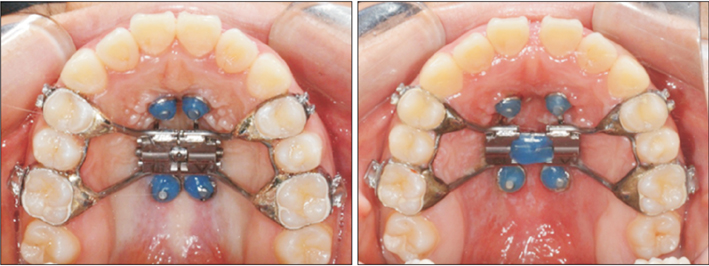
Figure 1 Miniscrew-assisted rapid palatal expander. Left, before expansion; right, after expansion.
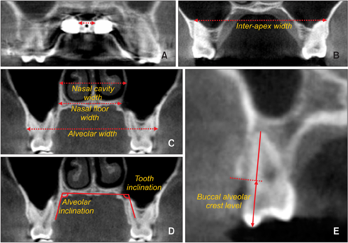
Figure 2 Measurements on the coronal images. A, Appliance expansion; B, interapex width; C, nasal cavity width (top), nasal floor width (middle), and alveolar width (bottom); D, alveolar inclination (left) and tooth inclination (right); E, buccal alveolar crest level.
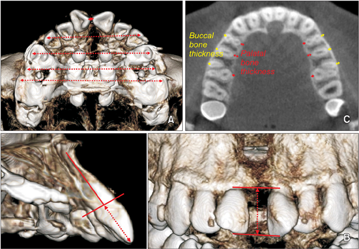
Figure 3 Measurements on three-dimensional and axial images. A, Intercusp width. From top to bottom, intercusp widths of the central incisors, canines, first premolars, second premolars, and first molars. B, Interproximal alveolar crest level (dashed arrow) between the central incisors; C, buccal and palatal bone thicknesses.
Cited by 4 articles
-
Stability of bimaxillary surgery involving intraoral vertical ramus osteotomy with or without presurgical miniscrew-assisted rapid palatal expansion in adult patients with skeletal Class III malocclusion
Yoon-Soo Ahn, Sung-Hwan Choi, Kee-Joon Lee, Young-Soo Jung, Hyoung-Seon Baik, Hyung-Seog Yu
Korean J Orthod. 2020;50(5):304-313. doi: 10.4041/kjod.2020.50.5.304.Dentofacial transverse development in Koreans according to skeletal maturation: A cross-sectional study
Soonshin Hwang, Yoonjeong Noh, Yoon Jeong Choi, Chooryung Chung, Hye Sun Lee, Kyung-Ho Kim
Korean J Orthod. 2018;48(1):39-47. doi: 10.4041/kjod.2018.48.1.39.Midfacial soft tissue changes after maxillary expansion using micro-implant-supported maxillary skeletal expanders in young adults: A retrospective study
Hieu Nguyen, Jeong Won Shin, Hai-Van Giap, Ki Beom Kim, Hwa Sung Chae, Young Ho Kim, Hae Won Choi
Korean J Orthod. 2021;51(3):145-156. doi: 10.4041/kjod.2021.51.3.145.Effectiveness of miniscrew assisted rapid palatal expansion using cone beam computed tomography: A systematic review and meta-analysis
Patchaya Siddhisaributr, Kornkanok Khlongwanitchakul, Niwat Anuwongnukroh, Somchai Manopatanakul, Nita Viwattanatipa
Korean J Orthod. 2022;52(3):182-200. doi: 10.4041/kjod21.256.
Reference
-
1. Melsen B. Palatal growth studied on human autopsy material. A histologic microradiographic study. Am J Orthod. 1975; 68:42–54.2. Kwak KH, Kim SS, Kim YI, Kim YD. Quantitative evaluation of midpalatal suture maturation via fractal analysis. Korean J Orthod. 2016; 46:323–330.
Article3. Jang HI, Kim SC, Chae JM, Kang KH, Cho JW, Chang NY, et al. Relationship between maturation indices and morphology of the midpalatal suture obtained using cone-beam computed tomography images. Korean J Orthod. 2016; 46:345–355.
Article4. Starnbach H, Bayne D, Cleall J, Subtelny JD. Facioskeletal and dental changes resulting from rapid maxillary expansion. Angle Orthod. 1966; 36:152–164.5. Wertz RA. Skeletal and dental changes accompanying rapid midpalatal suture opening. Am J Orthod. 1970; 58:41–66.
Article6. Bishara SE, Staley RN. Maxillary expansion: clinical implications. Am J Orthod Dentofacial Orthop. 1987; 91:3–14.
Article7. Björk A, Skieller V. Growth in width of the maxilla studied by the implant method. Scand J Plast Reconstr Surg. 1974; 8:26–33.
Article8. Suri L, Taneja P. Surgically assisted rapid palatal expansion: a literature review. Am J Orthod Dentofacial Orthop. 2008; 133:290–302.
Article9. Gauthier C, Voyer R, Paquette M, Rompré P, Papadakis A. Periodontal effects of surgically assisted rapid palatal expansion evaluated clinically and with cone-beam computerized tomography: 6-month preliminary results. Am J Orthod Dentofacial Orthop. 2011; 139:4 Suppl. S117–S128.
Article10. Lee KJ, Park YC, Park JY, Hwang WS. Miniscrew+assisted nonsurgical palatal expansion before orthognathic surgery for a patient with severe mandibular prognathism. Am J Orthod Dentofacial Orthop. 2010; 137:830–839.
Article11. Choi SH, Shi KK, Cha JY, Park YC, Lee KJ. Nonsurgical miniscrew-assisted rapid maxillary expansion results in acceptable stability in young adults. Angle Orthod. 2016; 86:713–720.
Article12. Park JJ, Park YC, Lee KJ, Cha JY, Tahk JH, Choi YJ. Skeletal and dentoalveolar changes after miniscrew-assisted rapid palatal expansion in young adults: a cone-beam computed tomography study. Korean J Orthod. 2017; 47:77–86.
Article13. Garib DG, Henriques JF, Janson G, de Freitas MR, Fernandes AY. Periodontal effects of rapid maxillary expansion with tooth-tissue-borne and tooth-borne expanders: a computed tomography evaluation. Am J Orthod Dentofacial Orthop. 2006; 129:749–758.
Article14. Rungcharassaeng K, Caruso JM, Kan JY, Kim J, Taylor G. Factors affecting buccal bone changes of maxillary posterior teeth after rapid maxillary expansion. Am J Orthod Dentofacial Orthop. 2007; 132:428.e1–428.e8.
Article15. Lin L, Ahn HW, Kim SJ, Moon SC, Kim SH, Nelson G. Tooth-borne vs bone-borne rapid maxillary expanders in late adolescence. Angle Orthod. 2015; 85:253–262.
Article16. Greenbaum KR, Zachrisson BU. The effect of palatal expansion therapy on the periodontal supporting tissues. Am J Orthod. 1982; 81:12–21.
Article17. Sarikaya S, Haydar B, Ciğer S, Ariyürek M. Changes in alveolar bone thickness due to retraction of anterior teeth. Am J Orthod Dentofacial Orthop. 2002; 122:15–26.
Article18. Choi YJ, Kim KH, Lee KJ, Chung CJ, Park YC. Radiographic evaluations of molar intrusion and changes with or without retention in rats. Angle Orthod. 2011; 81:389–396.
Article19. Vanarsdall RL Jr. Transverse dimension and long-term stability. Semin Orthod. 1999; 5:171–180.
Article20. Baccetti T, Franchi L, Toth LR, McNamara JA Jr. Treatment timing for Twin-block therapy. Am J Orthod Dentofacial Orthop. 2000; 118:159–170.
Article21. Kartalian A, Gohl E, Adamian M, Enciso R. Cone-beam computerized tomography evaluation of the maxillary dentoskeletal complex after rapid palatal expansion. Am J Orthod Dentofacial Orthop. 2010; 138:486–492.
Article22. Asscherickx K, Govaerts E, Aerts J, Vande Vannet B. Maxillary changes with bone-borne surgically assisted rapid palatal expansion: a prospective study. Am J Orthod Dentofacial Orthop. 2016; 149:374–383.
Article23. Gunyuz Toklu M, Germec-Cakan D, Tozlu M. Periodontal, dentoalveolar, and skeletal effects of tooth-borne and tooth-bone-borne expansion appliances. Am J Orthod Dentofacial Orthop. 2015; 148:97–109.
Article24. Persson M, Thilander B. Palatal suture closure in man from 15 to 35 years of age. Am J Orthod. 1977; 72:42–52.
Article25. Wehrbein H, Yildizhan F. The mid-palatal suture in young adults. A radiological-histological investigation. Eur J Orthod. 2001; 23:105–114.
Article26. Goldenberg DC, Goldenberg FC, Alonso N, Gebrin ES, Amaral TS, Scanavini MA, et al. Hyrax appliance opening and pattern of skeletal maxillary expansion after surgically assisted rapid palatal expansion: a computed tomography evaluation. Oral Surg Oral Med Oral Pathol Oral Radiol Endod. 2008; 106:812–819.
Article27. Zandi M, Miresmaeili A, Heidari A. Short-term skeletal and dental changes following bone-borne versus tooth-borne surgically assisted rapid maxillary expansion: a randomized clinical trial study. J Craniomaxillofac Surg. 2014; 42:1190–1195.
Article28. Isaacson RJ, Ingram AH. Forces produced by rapid maxillary expansion. II. Forces present during treatment. Angle Orthod. 1964; 34:261–270.29. Storey E. Tissue response to the movement of bones. Am J Orthod. 1973; 64:229–247.
Article30. Choi YJ, Kim KH, Lee KJ, Chung CJ, Park YC. Histomorphometric evaluation of maxillary molar roots and surrounding periodontium following molar intrusion in rats. Orthod Craniofac Res. 2015; 18:12–20.
Article31. Wehrbein H, Fuhrmann RA, Diedrich PR. Periodontal conditions after facial root tipping and palatal root torque of incisors. Am J Orthod Dentofacial Orthop. 1994; 106:455–462.
Article32. Grzesik WJ, Narayanan AS. Cementum and periodontal wound healing and regeneration. Crit Rev Oral Biol Med. 2002; 13:474–484.33. Kanzaki R, Daimaruya T, Takahashi I, Mitani H, Sugawara J. Remodeling of alveolar bone crest after molar intrusion with skeletal anchorage system in dogs. Am J Orthod Dentofacial Orthop. 2007; 131:343–351.
Article34. Angeletti P, Pereira MD, Gomes HC, Hino CT, Ferreira LM. Effect of low-level laser therapy (GaAlAs) on bone regeneration in midpalatal anterior suture after surgically assisted rapid maxillary expansion. Oral Surg Oral Med Oral Pathol Oral Radiol Endod. 2010; 109:e38–e46.
Article35. Wennström JL, Lindhe J, Sinclair F, Thilander B. Some periodontal tissue reactions to orthodontic tooth movement in monkeys. J Clin Periodontol. 1987; 14:121–129.
Article
- Full Text Links
-
- Actions
-
Cited
- CITED
-
- Close
- Share
-
- Similar articles
-
- Effectiveness of miniscrew assisted rapid palatal expansion using cone beam computed tomography: A systematic review and meta-analysis
- Effect and stability of miniscrew-assisted rapid palatal expansion: A systematic review and meta-analysis
- Stability of bimaxillary surgery involving intraoral vertical ramus osteotomy with or without presurgical miniscrew-assisted rapid palatal expansion in adult patients with skeletal Class III malocclusion
- Skeletal and dentoalveolar changes after miniscrew-assisted rapid palatal expansion in young adults: A cone-beam computed tomography study
- Skeletal and dentoalveolar effects of different types of microimplant-assisted rapid palatal expansion
