The esthetic prosthodontic treatments in maxillary anterior area, considering the gingival margin
- Affiliations
-
- 1Department of Prosthodontics, School of Dentistry, Kyung Hee University, Seoul, Republic of Korea. odontopia@khu.ac.kr
- KMID: 2388224
- DOI: http://doi.org/10.4047/jkap.2016.54.4.438
Abstract
- To enhance the esthetic appearance, the maxillary anterior area is important. It is possible to improve the esthetic appearance through the treatment of maxillary anterior area, which includes altering the color, form, and arrangement of teeth. When planning these treatments, clinicians should individualize personal demands, by using the information obtained from facial, dento-labial, dental, and gingival analysis. It is essential to properly prepare the gingival structure, which includes the height of gingival margin, the location of zenith, reconstruction of the interdental papillae, emergence profile, and symmetry. Clinicians often face unfavorable condition of the gingiva and the edentulous ridge, and appropriate management of the gingival structure is needed. In this case report, the patients were treated to improve the gingival conditions surrounding maxillary anterior teeth. By using conservative treatment without surgical intervention, such as application of pink porcelain, subgingival contour modelling and modification of pontic base, satisfactory esthetic results were gained.
Keyword
MeSH Terms
Figure
-
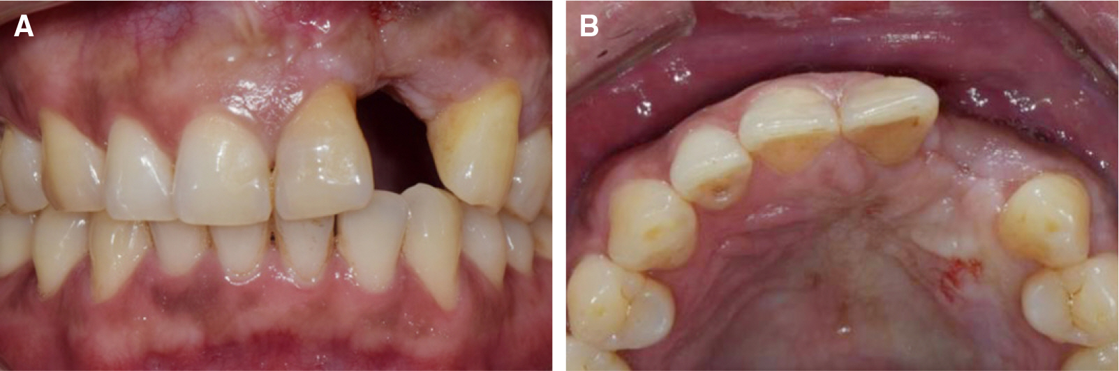
Fig. 1. Initial view. (A) Frontal view - vertical atrophy of ridge and gingival recession of left central incisor and canine, (B) Transverse view - slightly horizontal atrophy of ridge.
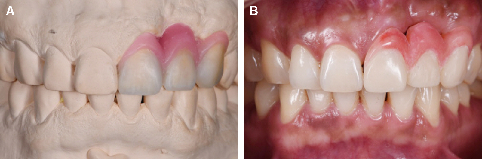
Fig. 2. (A) Diagnostic wax-up - mimics the gingival architecture, (B) Provisional restoration - transfer of diagnostic wax-up.
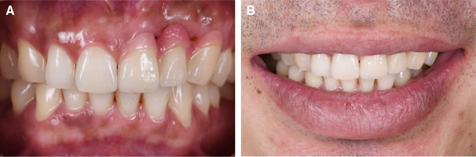
Fig. 3. (A) Definitive restoration - regain symmetry of gingival margin, (B) Evaluation of smile line.
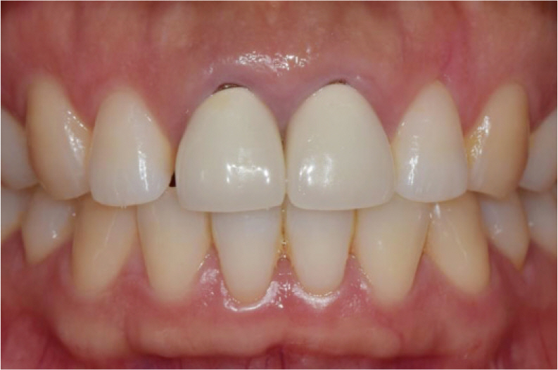
Fig. 4. Initial view. - unesthetic crown form and color, improper location of zeniths and distal space of right central incisor.
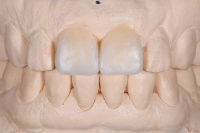
Fig. 5. Diagnostic wax-up - change of zenith and gingival margin of crowns.

Fig. 6. (A) Provisional restoration - improvement of zeniths and closure of distal space of right central incisor, (B) Completion of subgingival contour molding.
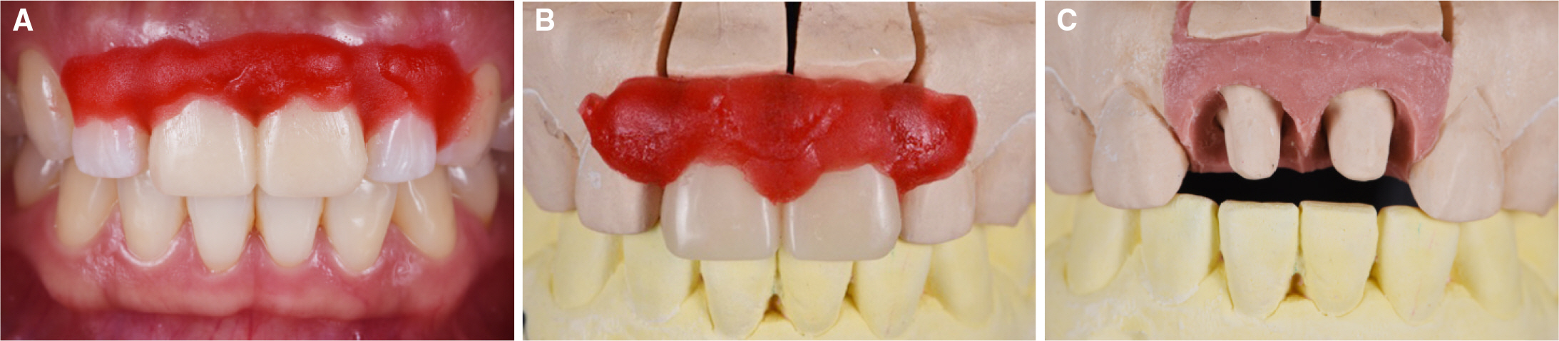
Fig. 7. Trasfer of gingival morphology. (A) Gingival impression with provisional restoration intraorally, (B) Acrylic resin zig and provisional restoration are seated on master cast, (C) After injection of silicone impression material.
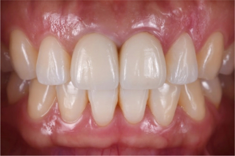
Fig. 8. Definitive restoration.
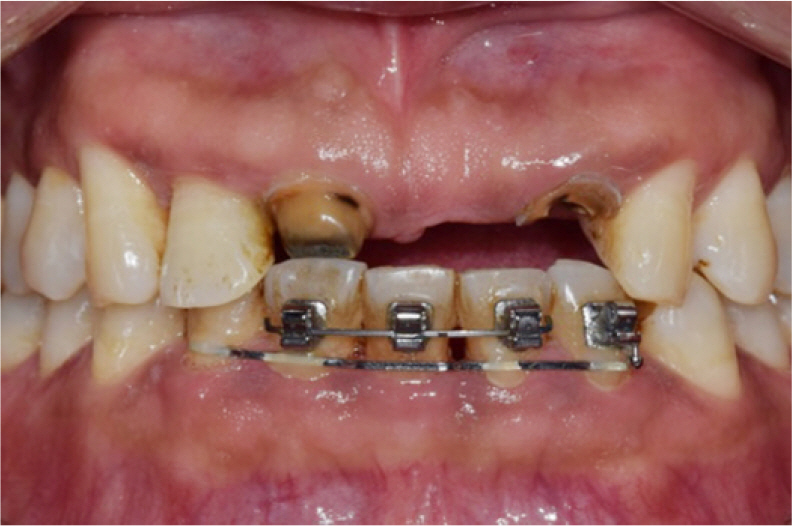
Fig. 9. Initial view.
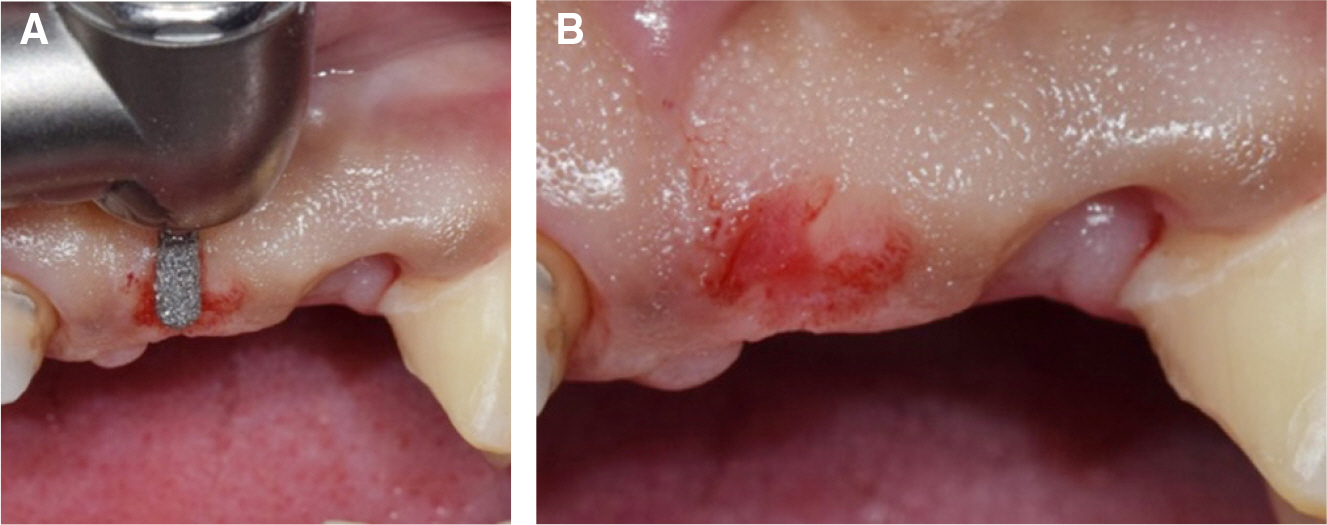
Fig. 10. Preparation of the gingiva under pontic base. (A) Shaping of the gingiva under the pontic base using high-speed handpiece, (B) Right after shaping the gingiva.

Fig. 11. (A) Preparation of retainer teeth and gingival shape - completely healed soft tissue under the area of pontic base, (B) Completion of provisional restoration - frontal view, (C) Completion of provisional restoration - lateral view. spontaneous emergence profile of pontics.
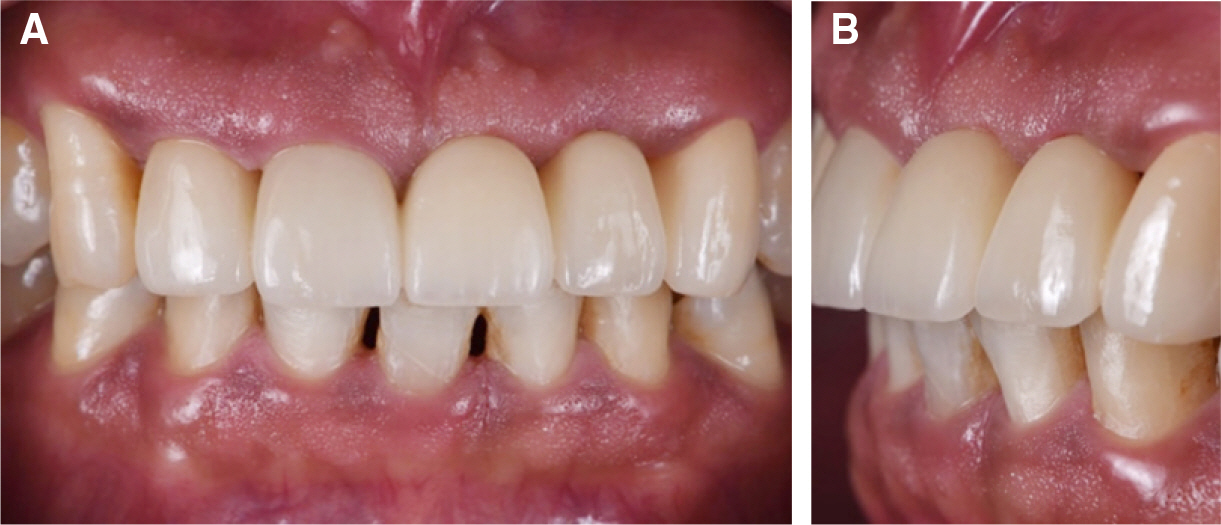
Fig. 12. Definitive restoration. (A) Frontal view, (B) Lateral view.
Reference
-
1.Fradeani M. Esthetic rehabilitation in fixed prosthodontics. 1:Esthetic analysis - a systematic approach to prosthetic treatment. Choi DG, Woo YH, Lee SB, Kwon KR, Paik J, Kim HS, editors. Seoul: DaehanNarae Publishing Inc.;2007.2.Magne P., Belser U. Bonded porcelain restorations in the anterior dentition. A biomimetic approach. Carol Stream; IL: Quintesence;2002. p. 58–64.3.Prichard J. Gingivoplasty, Gingivectomy, and osseous surgery. J Periodontal. 1961. 32:275–82.
Article4.Rufenacht CR. Fundamentals of esthetics. Chicago: Quintessence;1990. p. 67–134.5.Chiche GJ., Kokich VG., Caudill R. Diagnosis and treatment planning of esthetic problems. In: Chiche GJ, Pinault A (eds). Esthetics of anterior fixed prosthodontics. Chicago: Quintessence;1994. p. 33–52.6.Behrend DA. The design of multiple pontics. J Prosthet Dent. 1981. 46:634–8.
Article7.Haj-Ali R., Walker MP. A provisional fixed partial denture that simulates gingival tissue at the pontic-site defect. J Prosthodont. 2002. 11:46–8.8.Coachman C., Salama M., Garber D., Calamita M., Salama H., Cabral G. Prosthetic gingival reconstruction in fixed partial restorations. Part 3: laboratory procedures and maintenance. Int J Periodontics Restorative Dent. 2010. 30:19–29.9.Chu SJ., Tan JH., Stappert CF., Tarnow DP. Gingival zenith positions and levels of the maxillary anterior dentition. J Esthet Restor Dent. 2009. 21:113–20.
Article10.Noh K., Kwon KR., Kim HS., Kim DS., Pae A. Accurate transfer of soft tissue morphology with interim prosthesis to definitive cast. J Prosthet Dent. 2014. 111:159–62.11.Abrams L. Augmentation of the deformed residual edentulous ridge for fixed prosthesis. Compend Contin Educ Gen Dent. 1980. 1:205–13.12.Liu CL. Use of a modified ovate pontic in areas of ridge defects: a report of two cases. J Esthet Restor Dent. 2004. 16:273–81.
Article
- Full Text Links
-
- Actions
-
Cited
- CITED
-
- Close
- Share
-
- Similar articles
-
- Anterior esthetic restoration using DSD (digital smile design) for a patient with congenital missing tooth of maxillary central incisor
- Implant esthetic restoration with bone graft in the extended maxillary anterior area: A case report
- Esthetic improvements through systematic diagnosis and treatment procedures in patients with unesthetic maxillary anterior teeth proportion after orthodontic treatment: Case report
- Perception of maxillary anterior esthetics by dental professionals and lay people and topographical tooth-gingiva interface
- Single implant restoration with esthetic prosthodontic treatment in maxillary anterior tooth: A case report
