Investig Magn Reson Imaging.
2017 Jun;21(2):106-108. 10.13104/imri.2017.21.2.106.
Hyperperfusion in DWI Abnormality in a Patient with Acute Symptomatic Hypoglycemic Encephalopathy
- Affiliations
-
- 1Department of Radiology, Pohang Stroke and Spine Hospital, Pohang, Korea. jkcontrast@naver.com
- KMID: 2385608
- DOI: http://doi.org/10.13104/imri.2017.21.2.106
Abstract
- The perfusion change in acute symptomatic hypoglycemic encephalopathy (ASHE) is not well known. We present the perfusion-weighted imaging of a patient with ASHE. The area of diffusion-weighted imaging abnormalities and adjacent normal-appearing white matter showed increased cerebral blood volume and flow, and shortening of time-to-peak.
Figure
-
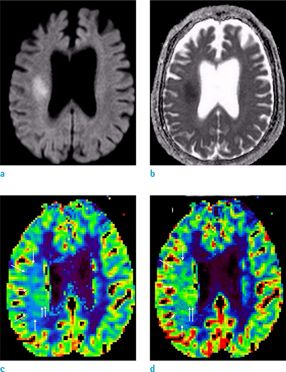
Fig. 1 Diffusion-weighted imaging (DWI) (a) and apparent diffusion coefficient map (b) revealed diffusion restriction along the right corona radiata of the corticospinal tract. Relative cerebral blood volume (c) and relative cerebral blood flow (d) showed increased perfusion in the area of DWI abnormalities and adjacent normal-appearing white matter. Hyperperfusion was predominant in the DWI abnormalities and the deep white matter posterior to the DWI lesion (double arrows), and the white matter near to the cortex (arrows) showed mildly increased perfusion.
Reference
-
1. Yong AW, Morris Z, Shuler K, Smith C, Wardlaw J. Acute symptomatic hypoglycaemia mimicking ischaemic stroke on imaging: a systemic review. BMC Neurol. 2012; 12:139.2. Tallroth G, Ryding E, Agardh CD. Regional cerebral blood flow in normal man during insulin-induced hypoglycemia and in the recovery period following glucose infusion. Metabolism. 1992; 41:717–721.3. Cordonnier C, Oppenheim C, Lamy C, Meder JF, Mas JL. Serial diffusion and perfusion-weighted MR in transient hypoglycemia. Neurology. 2005; 65:175.4. Bottcher J, Kunze A, Kurrat C, et al. Localized reversible reduction of apparent diffusion coefficient in transient hypoglycemia-induced hemiparesis. Stroke. 2005; 36:e20–e22.5. Lo L, Tan AC, Umapathi T, Lim CC. Diffusion-weighted MR imaging in early diagnosis and prognosis of hypoglycemia. AJNR Am J Neuroradiol. 2006; 27:1222–1224.6. Aoki T, Sato T, Hasegawa K, Ishizaki R, Saiki M. Reversible hyperintensity lesion on diffusion-weighted MRI in hypoglycemic coma. Neurology. 2004; 63:392–393.7. Kim JH, Lim MK, Jeon TY, et al. Diffusion and perfusion characteristics of MELAS (mitochondrial myopathy, encephalopathy, lactic acidosis, and stroke-like episode) in thirteen patients. Korean J Radiol. 2011; 12:15–24.8. Li R, Xiao HF, Lyu JH, JJ Wang D, Ma L, Lou X. Differential diagnosis of mitochondrial encephalopathy with lactic acidosis and stroke-like episodes (MELAS) and ischemic stroke using 3D pseudocontinuous arterial spin labeling. J Magn Reson Imaging. 2017; 45:199–206.
- Full Text Links
-
- Actions
-
Cited
- CITED
-
- Close
- Share
-
- Similar articles
-
- Rapid Regression of White Matter Changes in Hypoglycemic Encephalopathy
- A Case of Hypoglycemic Encephalopathy with Lesion in the Hippocampus on Diffusion-Weighted MRI
- Acute Hyperammonemic Encephalopathy with Features on Diffusion-Weighted Images: Report of Two Cases
- Hypoglycemic Encephalopathy with Reversible Unilateral Hippocampal Lesion on Brain MRI
- Consideration of Prognostic Factors in Hypoglycemic Encephalopathy
